US7115558B2 - P450RAI-2(P450 Cytochrome 26B), encoding nucleic acid molecules and methods and uses thereof - Google Patents
P450RAI-2(P450 Cytochrome 26B), encoding nucleic acid molecules and methods and uses thereof Download PDFInfo
- Publication number
- US7115558B2 US7115558B2 US10/433,485 US43348503A US7115558B2 US 7115558 B2 US7115558 B2 US 7115558B2 US 43348503 A US43348503 A US 43348503A US 7115558 B2 US7115558 B2 US 7115558B2
- Authority
- US
- United States
- Prior art keywords
- p450rai
- protein
- cells
- expression
- seq
- Prior art date
- Legal status (The legal status is an assumption and is not a legal conclusion. Google has not performed a legal analysis and makes no representation as to the accuracy of the status listed.)
- Expired - Fee Related, expires
Links
- 238000000034 method Methods 0.000 title abstract description 151
- 102100039281 Cytochrome P450 26B1 Human genes 0.000 title abstract description 26
- 101000745883 Homo sapiens Cytochrome P450 26B1 Proteins 0.000 title abstract description 5
- 150000007523 nucleic acids Chemical class 0.000 title description 177
- 102000039446 nucleic acids Human genes 0.000 title description 145
- 108020004707 nucleic acids Proteins 0.000 title description 145
- 102000018832 Cytochromes Human genes 0.000 title description 11
- 108010052832 Cytochromes Proteins 0.000 title description 11
- 108010015742 Cytochrome P-450 Enzyme System Proteins 0.000 claims abstract description 21
- 108090000623 proteins and genes Proteins 0.000 claims description 365
- 229930002330 retinoic acid Natural products 0.000 claims description 315
- SHGAZHPCJJPHSC-YCNIQYBTSA-N all-trans-retinoic acid Chemical compound OC(=O)\C=C(/C)\C=C\C=C(/C)\C=C\C1=C(C)CCCC1(C)C SHGAZHPCJJPHSC-YCNIQYBTSA-N 0.000 claims description 311
- 102000004169 proteins and genes Human genes 0.000 claims description 293
- 229960001727 tretinoin Drugs 0.000 claims description 195
- 125000003275 alpha amino acid group Chemical group 0.000 claims description 48
- 102000002004 Cytochrome P-450 Enzyme System Human genes 0.000 claims description 20
- 230000004044 response Effects 0.000 claims description 9
- 230000002163 immunogen Effects 0.000 claims description 8
- 210000004027 cell Anatomy 0.000 abstract description 277
- 241000282414 Homo sapiens Species 0.000 abstract description 120
- 239000012634 fragment Substances 0.000 abstract description 58
- 150000001413 amino acids Chemical class 0.000 abstract description 41
- 108090000765 processed proteins & peptides Proteins 0.000 abstract description 40
- 208000037265 diseases, disorders, signs and symptoms Diseases 0.000 abstract description 38
- 206010028980 Neoplasm Diseases 0.000 abstract description 34
- 239000002207 metabolite Substances 0.000 abstract description 34
- 238000011282 treatment Methods 0.000 abstract description 31
- 239000003112 inhibitor Substances 0.000 abstract description 23
- 102000004196 processed proteins & peptides Human genes 0.000 abstract description 21
- 210000001638 cerebellum Anatomy 0.000 abstract description 17
- 101100007328 Cocos nucifera COS-1 gene Proteins 0.000 abstract description 16
- 230000001939 inductive effect Effects 0.000 abstract description 16
- 210000004072 lung Anatomy 0.000 abstract description 16
- 208000035475 disorder Diseases 0.000 abstract description 14
- 201000011510 cancer Diseases 0.000 abstract description 13
- 210000004556 brain Anatomy 0.000 abstract description 12
- 238000006243 chemical reaction Methods 0.000 abstract description 12
- 229920001184 polypeptide Polymers 0.000 abstract description 12
- 206010006187 Breast cancer Diseases 0.000 abstract description 8
- 208000026310 Breast neoplasm Diseases 0.000 abstract description 8
- 206010058467 Lung neoplasm malignant Diseases 0.000 abstract description 8
- 201000005202 lung cancer Diseases 0.000 abstract description 8
- 208000020816 lung neoplasm Diseases 0.000 abstract description 8
- 102000040430 polynucleotide Human genes 0.000 abstract description 6
- 108091033319 polynucleotide Proteins 0.000 abstract description 6
- 239000002157 polynucleotide Substances 0.000 abstract description 6
- 210000004748 cultured cell Anatomy 0.000 abstract description 5
- 208000014644 Brain disease Diseases 0.000 abstract 1
- 102000003849 Cytochrome P450 Human genes 0.000 abstract 1
- 235000018102 proteins Nutrition 0.000 description 287
- 230000014509 gene expression Effects 0.000 description 181
- 239000000523 sample Substances 0.000 description 125
- YVOOPGWEIRIUOX-UHFFFAOYSA-N 2-azanyl-3-sulfanyl-propanoic acid Chemical compound SCC(N)C(O)=O.SCC(N)C(O)=O YVOOPGWEIRIUOX-UHFFFAOYSA-N 0.000 description 105
- 230000000694 effects Effects 0.000 description 105
- 125000003729 nucleotide group Chemical group 0.000 description 98
- 239000002773 nucleotide Substances 0.000 description 84
- 241001465754 Metazoa Species 0.000 description 70
- 241000699666 Mus <mouse, genus> Species 0.000 description 70
- 210000001519 tissue Anatomy 0.000 description 68
- 230000004060 metabolic process Effects 0.000 description 66
- 108020004414 DNA Proteins 0.000 description 61
- 108091032973 (ribonucleotides)n+m Proteins 0.000 description 58
- 150000004492 retinoid derivatives Chemical class 0.000 description 55
- 239000000758 substrate Substances 0.000 description 51
- 235000001014 amino acid Nutrition 0.000 description 47
- 239000002299 complementary DNA Substances 0.000 description 45
- 229940024606 amino acid Drugs 0.000 description 43
- 239000003795 chemical substances by application Substances 0.000 description 42
- 230000000692 anti-sense effect Effects 0.000 description 38
- 239000013615 primer Substances 0.000 description 37
- 210000002257 embryonic structure Anatomy 0.000 description 36
- 238000009396 hybridization Methods 0.000 description 35
- FPIPGXGPPPQFEQ-OVSJKPMPSA-N all-trans-retinol Chemical compound OC\C=C(/C)\C=C\C=C(/C)\C=C\C1=C(C)CCCC1(C)C FPIPGXGPPPQFEQ-OVSJKPMPSA-N 0.000 description 34
- 108091028043 Nucleic acid sequence Proteins 0.000 description 33
- 150000001875 compounds Chemical class 0.000 description 33
- 239000000047 product Substances 0.000 description 33
- 102000004190 Enzymes Human genes 0.000 description 31
- 108090000790 Enzymes Proteins 0.000 description 31
- 229940088598 enzyme Drugs 0.000 description 31
- 238000012216 screening Methods 0.000 description 31
- 230000009261 transgenic effect Effects 0.000 description 31
- 239000003814 drug Substances 0.000 description 30
- 238000003752 polymerase chain reaction Methods 0.000 description 29
- 108700008625 Reporter Genes Proteins 0.000 description 26
- 229940079593 drug Drugs 0.000 description 26
- 239000013604 expression vector Substances 0.000 description 25
- 201000010099 disease Diseases 0.000 description 24
- 230000027455 binding Effects 0.000 description 23
- 239000000203 mixture Substances 0.000 description 23
- 230000001105 regulatory effect Effects 0.000 description 23
- 230000006698 induction Effects 0.000 description 22
- 239000000126 substance Substances 0.000 description 22
- 239000013598 vector Substances 0.000 description 22
- 239000000243 solution Substances 0.000 description 21
- 241000252212 Danio rerio Species 0.000 description 20
- 108091034117 Oligonucleotide Proteins 0.000 description 20
- 230000001965 increasing effect Effects 0.000 description 20
- 238000010186 staining Methods 0.000 description 20
- FPIPGXGPPPQFEQ-UHFFFAOYSA-N 13-cis retinol Natural products OCC=C(C)C=CC=C(C)C=CC1=C(C)CCCC1(C)C FPIPGXGPPPQFEQ-UHFFFAOYSA-N 0.000 description 19
- 238000000636 Northern blotting Methods 0.000 description 17
- 238000004458 analytical method Methods 0.000 description 16
- 230000000295 complement effect Effects 0.000 description 16
- 239000003550 marker Substances 0.000 description 16
- 238000003757 reverse transcription PCR Methods 0.000 description 16
- 238000006467 substitution reaction Methods 0.000 description 16
- QTBSBXVTEAMEQO-UHFFFAOYSA-N Acetic acid Chemical compound CC(O)=O QTBSBXVTEAMEQO-UHFFFAOYSA-N 0.000 description 15
- WEVYAHXRMPXWCK-UHFFFAOYSA-N Acetonitrile Chemical compound CC#N WEVYAHXRMPXWCK-UHFFFAOYSA-N 0.000 description 15
- 101100114674 Homo sapiens CYP26B1 gene Proteins 0.000 description 15
- 238000004519 manufacturing process Methods 0.000 description 15
- 230000002285 radioactive effect Effects 0.000 description 15
- 238000012360 testing method Methods 0.000 description 15
- IAZDPXIOMUYVGZ-UHFFFAOYSA-N Dimethylsulphoxide Chemical compound CS(C)=O IAZDPXIOMUYVGZ-UHFFFAOYSA-N 0.000 description 14
- 238000004128 high performance liquid chromatography Methods 0.000 description 14
- 239000000463 material Substances 0.000 description 14
- 229960003471 retinol Drugs 0.000 description 14
- 235000020944 retinol Nutrition 0.000 description 14
- 239000011607 retinol Substances 0.000 description 14
- XLYOFNOQVPJJNP-UHFFFAOYSA-N water Chemical compound O XLYOFNOQVPJJNP-UHFFFAOYSA-N 0.000 description 14
- -1 RA glucuronide Chemical class 0.000 description 13
- 241000700159 Rattus Species 0.000 description 13
- 238000003556 assay Methods 0.000 description 13
- 239000003153 chemical reaction reagent Substances 0.000 description 13
- 108020001507 fusion proteins Proteins 0.000 description 13
- 102000037865 fusion proteins Human genes 0.000 description 13
- 238000003259 recombinant expression Methods 0.000 description 13
- 238000005406 washing Methods 0.000 description 13
- 101150053185 P450 gene Proteins 0.000 description 12
- PSQYTAPXSHCGMF-BQYQJAHWSA-N beta-ionone group Chemical group CC1=C(C(CCC1)(C)C)/C=C/C(C)=O PSQYTAPXSHCGMF-BQYQJAHWSA-N 0.000 description 12
- 230000015572 biosynthetic process Effects 0.000 description 12
- 230000015556 catabolic process Effects 0.000 description 12
- 229960001760 dimethyl sulfoxide Drugs 0.000 description 12
- 239000000499 gel Substances 0.000 description 12
- 102000006602 glyceraldehyde-3-phosphate dehydrogenase Human genes 0.000 description 12
- 108020004445 glyceraldehyde-3-phosphate dehydrogenase Proteins 0.000 description 12
- 230000002401 inhibitory effect Effects 0.000 description 12
- 239000002609 medium Substances 0.000 description 12
- 210000000952 spleen Anatomy 0.000 description 12
- SHGAZHPCJJPHSC-NUEINMDLSA-N Isotretinoin Chemical compound OC(=O)C=C(C)/C=C/C=C(C)C=CC1=C(C)CCCC1(C)C SHGAZHPCJJPHSC-NUEINMDLSA-N 0.000 description 11
- 210000004408 hybridoma Anatomy 0.000 description 11
- 238000001727 in vivo Methods 0.000 description 11
- 230000035772 mutation Effects 0.000 description 11
- 210000001525 retina Anatomy 0.000 description 11
- 235000020945 retinal Nutrition 0.000 description 11
- 239000011604 retinal Substances 0.000 description 11
- 238000010561 standard procedure Methods 0.000 description 11
- NCYCYZXNIZJOKI-UHFFFAOYSA-N vitamin A aldehyde Natural products O=CC=C(C)C=CC=C(C)C=CC1=C(C)CCCC1(C)C NCYCYZXNIZJOKI-UHFFFAOYSA-N 0.000 description 11
- 108091026890 Coding region Proteins 0.000 description 10
- 102100039282 Cytochrome P450 26A1 Human genes 0.000 description 10
- 108010022037 Retinoic Acid 4-Hydroxylase Proteins 0.000 description 10
- 108700019146 Transgenes Proteins 0.000 description 10
- 230000006907 apoptotic process Effects 0.000 description 10
- 238000011161 development Methods 0.000 description 10
- 230000018109 developmental process Effects 0.000 description 10
- 230000005764 inhibitory process Effects 0.000 description 10
- 230000002503 metabolic effect Effects 0.000 description 10
- 230000003647 oxidation Effects 0.000 description 10
- 238000007254 oxidation reaction Methods 0.000 description 10
- 230000001225 therapeutic effect Effects 0.000 description 10
- XMAYWYJOQHXEEK-OZXSUGGESA-N (2R,4S)-ketoconazole Chemical compound C1CN(C(=O)C)CCN1C(C=C1)=CC=C1OC[C@@H]1O[C@@](CN2C=NC=C2)(C=2C(=CC(Cl)=CC=2)Cl)OC1 XMAYWYJOQHXEEK-OZXSUGGESA-N 0.000 description 9
- SHGAZHPCJJPHSC-UHFFFAOYSA-N Panrexin Chemical compound OC(=O)C=C(C)C=CC=C(C)C=CC1=C(C)CCCC1(C)C SHGAZHPCJJPHSC-UHFFFAOYSA-N 0.000 description 9
- 208000037065 Subacute sclerosing leukoencephalitis Diseases 0.000 description 9
- 206010042297 Subacute sclerosing panencephalitis Diseases 0.000 description 9
- JLCPHMBAVCMARE-UHFFFAOYSA-N [3-[[3-[[3-[[3-[[3-[[3-[[3-[[3-[[3-[[3-[[3-[[5-(2-amino-6-oxo-1H-purin-9-yl)-3-[[3-[[3-[[3-[[3-[[3-[[5-(2-amino-6-oxo-1H-purin-9-yl)-3-[[5-(2-amino-6-oxo-1H-purin-9-yl)-3-hydroxyoxolan-2-yl]methoxy-hydroxyphosphoryl]oxyoxolan-2-yl]methoxy-hydroxyphosphoryl]oxy-5-(5-methyl-2,4-dioxopyrimidin-1-yl)oxolan-2-yl]methoxy-hydroxyphosphoryl]oxy-5-(6-aminopurin-9-yl)oxolan-2-yl]methoxy-hydroxyphosphoryl]oxy-5-(6-aminopurin-9-yl)oxolan-2-yl]methoxy-hydroxyphosphoryl]oxy-5-(6-aminopurin-9-yl)oxolan-2-yl]methoxy-hydroxyphosphoryl]oxy-5-(6-aminopurin-9-yl)oxolan-2-yl]methoxy-hydroxyphosphoryl]oxyoxolan-2-yl]methoxy-hydroxyphosphoryl]oxy-5-(5-methyl-2,4-dioxopyrimidin-1-yl)oxolan-2-yl]methoxy-hydroxyphosphoryl]oxy-5-(4-amino-2-oxopyrimidin-1-yl)oxolan-2-yl]methoxy-hydroxyphosphoryl]oxy-5-(5-methyl-2,4-dioxopyrimidin-1-yl)oxolan-2-yl]methoxy-hydroxyphosphoryl]oxy-5-(5-methyl-2,4-dioxopyrimidin-1-yl)oxolan-2-yl]methoxy-hydroxyphosphoryl]oxy-5-(6-aminopurin-9-yl)oxolan-2-yl]methoxy-hydroxyphosphoryl]oxy-5-(6-aminopurin-9-yl)oxolan-2-yl]methoxy-hydroxyphosphoryl]oxy-5-(4-amino-2-oxopyrimidin-1-yl)oxolan-2-yl]methoxy-hydroxyphosphoryl]oxy-5-(4-amino-2-oxopyrimidin-1-yl)oxolan-2-yl]methoxy-hydroxyphosphoryl]oxy-5-(4-amino-2-oxopyrimidin-1-yl)oxolan-2-yl]methoxy-hydroxyphosphoryl]oxy-5-(6-aminopurin-9-yl)oxolan-2-yl]methoxy-hydroxyphosphoryl]oxy-5-(4-amino-2-oxopyrimidin-1-yl)oxolan-2-yl]methyl [5-(6-aminopurin-9-yl)-2-(hydroxymethyl)oxolan-3-yl] hydrogen phosphate Polymers Cc1cn(C2CC(OP(O)(=O)OCC3OC(CC3OP(O)(=O)OCC3OC(CC3O)n3cnc4c3nc(N)[nH]c4=O)n3cnc4c3nc(N)[nH]c4=O)C(COP(O)(=O)OC3CC(OC3COP(O)(=O)OC3CC(OC3COP(O)(=O)OC3CC(OC3COP(O)(=O)OC3CC(OC3COP(O)(=O)OC3CC(OC3COP(O)(=O)OC3CC(OC3COP(O)(=O)OC3CC(OC3COP(O)(=O)OC3CC(OC3COP(O)(=O)OC3CC(OC3COP(O)(=O)OC3CC(OC3COP(O)(=O)OC3CC(OC3COP(O)(=O)OC3CC(OC3COP(O)(=O)OC3CC(OC3COP(O)(=O)OC3CC(OC3COP(O)(=O)OC3CC(OC3COP(O)(=O)OC3CC(OC3COP(O)(=O)OC3CC(OC3CO)n3cnc4c(N)ncnc34)n3ccc(N)nc3=O)n3cnc4c(N)ncnc34)n3ccc(N)nc3=O)n3ccc(N)nc3=O)n3ccc(N)nc3=O)n3cnc4c(N)ncnc34)n3cnc4c(N)ncnc34)n3cc(C)c(=O)[nH]c3=O)n3cc(C)c(=O)[nH]c3=O)n3ccc(N)nc3=O)n3cc(C)c(=O)[nH]c3=O)n3cnc4c3nc(N)[nH]c4=O)n3cnc4c(N)ncnc34)n3cnc4c(N)ncnc34)n3cnc4c(N)ncnc34)n3cnc4c(N)ncnc34)O2)c(=O)[nH]c1=O JLCPHMBAVCMARE-UHFFFAOYSA-N 0.000 description 9
- 239000000284 extract Substances 0.000 description 9
- 210000005260 human cell Anatomy 0.000 description 9
- 238000005805 hydroxylation reaction Methods 0.000 description 9
- 238000011534 incubation Methods 0.000 description 9
- 229960004125 ketoconazole Drugs 0.000 description 9
- 238000002372 labelling Methods 0.000 description 9
- 210000003141 lower extremity Anatomy 0.000 description 9
- 230000001404 mediated effect Effects 0.000 description 9
- 238000012544 monitoring process Methods 0.000 description 9
- 239000013612 plasmid Substances 0.000 description 9
- 239000012679 serum free medium Substances 0.000 description 9
- 238000003786 synthesis reaction Methods 0.000 description 9
- 238000013518 transcription Methods 0.000 description 9
- 230000035897 transcription Effects 0.000 description 9
- 239000003981 vehicle Substances 0.000 description 9
- 108010035563 Chloramphenicol O-acetyltransferase Proteins 0.000 description 8
- 102000053602 DNA Human genes 0.000 description 8
- 239000011543 agarose gel Substances 0.000 description 8
- 230000033228 biological regulation Effects 0.000 description 8
- 238000007796 conventional method Methods 0.000 description 8
- 238000012217 deletion Methods 0.000 description 8
- 230000037430 deletion Effects 0.000 description 8
- 239000003623 enhancer Substances 0.000 description 8
- 210000003194 forelimb Anatomy 0.000 description 8
- UYTPUPDQBNUYGX-UHFFFAOYSA-N guanine Chemical compound O=C1NC(N)=NC2=C1N=CN2 UYTPUPDQBNUYGX-UHFFFAOYSA-N 0.000 description 8
- 239000012528 membrane Substances 0.000 description 8
- 230000002207 retinal effect Effects 0.000 description 8
- 241000894007 species Species 0.000 description 8
- 208000036762 Acute promyelocytic leukaemia Diseases 0.000 description 7
- 206010058314 Dysplasia Diseases 0.000 description 7
- PEDCQBHIVMGVHV-UHFFFAOYSA-N Glycerine Chemical compound OCC(O)CO PEDCQBHIVMGVHV-UHFFFAOYSA-N 0.000 description 7
- OKKJLVBELUTLKV-UHFFFAOYSA-N Methanol Chemical compound OC OKKJLVBELUTLKV-UHFFFAOYSA-N 0.000 description 7
- 241000699670 Mus sp. Species 0.000 description 7
- 208000033826 Promyelocytic Acute Leukemia Diseases 0.000 description 7
- GGCUJPCCTQNTJF-FRCNGJHJSA-N all-trans-4-oxoretinoic acid Chemical compound OC(=O)\C=C(/C)\C=C\C=C(/C)\C=C\C1=C(C)C(=O)CCC1(C)C GGCUJPCCTQNTJF-FRCNGJHJSA-N 0.000 description 7
- 230000003925 brain function Effects 0.000 description 7
- 230000001419 dependent effect Effects 0.000 description 7
- 230000004927 fusion Effects 0.000 description 7
- 230000033444 hydroxylation Effects 0.000 description 7
- 238000003780 insertion Methods 0.000 description 7
- 230000037431 insertion Effects 0.000 description 7
- 108020004999 messenger RNA Proteins 0.000 description 7
- 230000004048 modification Effects 0.000 description 7
- 238000012986 modification Methods 0.000 description 7
- 239000012071 phase Substances 0.000 description 7
- 238000002360 preparation method Methods 0.000 description 7
- 210000002966 serum Anatomy 0.000 description 7
- 108010085238 Actins Proteins 0.000 description 6
- 102000007469 Actins Human genes 0.000 description 6
- 239000006144 Dulbecco’s modified Eagle's medium Substances 0.000 description 6
- 108700024394 Exon Proteins 0.000 description 6
- 241000282412 Homo Species 0.000 description 6
- 241001529936 Murinae Species 0.000 description 6
- 206010035226 Plasma cell myeloma Diseases 0.000 description 6
- FAPWRFPIFSIZLT-UHFFFAOYSA-M Sodium chloride Chemical compound [Na+].[Cl-] FAPWRFPIFSIZLT-UHFFFAOYSA-M 0.000 description 6
- 241000269370 Xenopus <genus> Species 0.000 description 6
- 101001099854 Xenopus laevis Cellular retinoic acid-binding protein 2 Proteins 0.000 description 6
- 239000002253 acid Substances 0.000 description 6
- KGUMXGDKXYTTEY-FRCNGJHJSA-N all-trans-4-hydroxyretinoic acid Chemical compound OC(=O)\C=C(/C)\C=C\C=C(/C)\C=C\C1=C(C)C(O)CCC1(C)C KGUMXGDKXYTTEY-FRCNGJHJSA-N 0.000 description 6
- 125000000539 amino acid group Chemical group 0.000 description 6
- 239000002246 antineoplastic agent Substances 0.000 description 6
- 230000008901 benefit Effects 0.000 description 6
- 102000005936 beta-Galactosidase Human genes 0.000 description 6
- 108010005774 beta-Galactosidase Proteins 0.000 description 6
- 230000004071 biological effect Effects 0.000 description 6
- 210000005013 brain tissue Anatomy 0.000 description 6
- OPTASPLRGRRNAP-UHFFFAOYSA-N cytosine Chemical compound NC=1C=CNC(=O)N=1 OPTASPLRGRRNAP-UHFFFAOYSA-N 0.000 description 6
- 230000007423 decrease Effects 0.000 description 6
- LOKCTEFSRHRXRJ-UHFFFAOYSA-I dipotassium trisodium dihydrogen phosphate hydrogen phosphate dichloride Chemical compound P(=O)(O)(O)[O-].[K+].P(=O)(O)([O-])[O-].[Na+].[Na+].[Cl-].[K+].[Cl-].[Na+] LOKCTEFSRHRXRJ-UHFFFAOYSA-I 0.000 description 6
- 210000004602 germ cell Anatomy 0.000 description 6
- 201000000050 myeloid neoplasm Diseases 0.000 description 6
- 208000002154 non-small cell lung carcinoma Diseases 0.000 description 6
- 239000008194 pharmaceutical composition Substances 0.000 description 6
- 239000002953 phosphate buffered saline Substances 0.000 description 6
- 230000004952 protein activity Effects 0.000 description 6
- 230000002441 reversible effect Effects 0.000 description 6
- 231100000331 toxic Toxicity 0.000 description 6
- 230000002588 toxic effect Effects 0.000 description 6
- 238000013519 translation Methods 0.000 description 6
- 230000014616 translation Effects 0.000 description 6
- 210000004881 tumor cell Anatomy 0.000 description 6
- 238000011144 upstream manufacturing Methods 0.000 description 6
- 235000019155 vitamin A Nutrition 0.000 description 6
- 239000011719 vitamin A Substances 0.000 description 6
- 229940045997 vitamin a Drugs 0.000 description 6
- GFFGJBXGBJISGV-UHFFFAOYSA-N Adenine Chemical compound NC1=NC=NC2=C1N=CN2 GFFGJBXGBJISGV-UHFFFAOYSA-N 0.000 description 5
- 208000023275 Autoimmune disease Diseases 0.000 description 5
- 108020004705 Codon Proteins 0.000 description 5
- 108020004635 Complementary DNA Proteins 0.000 description 5
- 101710130824 Cytochrome P450 26B1 Proteins 0.000 description 5
- 101001100103 Homo sapiens Retinoic acid-induced protein 2 Proteins 0.000 description 5
- 108010029485 Protein Isoforms Proteins 0.000 description 5
- 102000001708 Protein Isoforms Human genes 0.000 description 5
- 201000004681 Psoriasis Diseases 0.000 description 5
- 108020004511 Recombinant DNA Proteins 0.000 description 5
- 102000007056 Recombinant Fusion Proteins Human genes 0.000 description 5
- 108010008281 Recombinant Fusion Proteins Proteins 0.000 description 5
- NCYCYZXNIZJOKI-OVSJKPMPSA-N Retinaldehyde Chemical compound O=C\C=C(/C)\C=C\C=C(/C)\C=C\C1=C(C)CCCC1(C)C NCYCYZXNIZJOKI-OVSJKPMPSA-N 0.000 description 5
- 102100038452 Retinoic acid-induced protein 2 Human genes 0.000 description 5
- FPIPGXGPPPQFEQ-BOOMUCAASA-N Vitamin A Natural products OC/C=C(/C)\C=C\C=C(\C)/C=C/C1=C(C)CCCC1(C)C FPIPGXGPPPQFEQ-BOOMUCAASA-N 0.000 description 5
- 230000009471 action Effects 0.000 description 5
- 239000000427 antigen Substances 0.000 description 5
- 108091007433 antigens Proteins 0.000 description 5
- 102000036639 antigens Human genes 0.000 description 5
- 230000001580 bacterial effect Effects 0.000 description 5
- 238000001514 detection method Methods 0.000 description 5
- 230000008034 disappearance Effects 0.000 description 5
- 239000003937 drug carrier Substances 0.000 description 5
- ZMMJGEGLRURXTF-UHFFFAOYSA-N ethidium bromide Chemical compound [Br-].C12=CC(N)=CC=C2C2=CC=C(N)C=C2[N+](CC)=C1C1=CC=CC=C1 ZMMJGEGLRURXTF-UHFFFAOYSA-N 0.000 description 5
- 229960005542 ethidium bromide Drugs 0.000 description 5
- 230000006870 function Effects 0.000 description 5
- 230000002068 genetic effect Effects 0.000 description 5
- 210000003734 kidney Anatomy 0.000 description 5
- 210000004185 liver Anatomy 0.000 description 5
- 210000004962 mammalian cell Anatomy 0.000 description 5
- 230000007246 mechanism Effects 0.000 description 5
- 210000004379 membrane Anatomy 0.000 description 5
- 210000001589 microsome Anatomy 0.000 description 5
- 230000007935 neutral effect Effects 0.000 description 5
- 238000000252 photodiode array detection Methods 0.000 description 5
- 230000008569 process Effects 0.000 description 5
- 238000003345 scintillation counting Methods 0.000 description 5
- 238000012546 transfer Methods 0.000 description 5
- YBJHBAHKTGYVGT-ZKWXMUAHSA-N (+)-Biotin Chemical compound N1C(=O)N[C@@H]2[C@H](CCCCC(=O)O)SC[C@@H]21 YBJHBAHKTGYVGT-ZKWXMUAHSA-N 0.000 description 4
- OIXLLKLZKCBCPS-RZVRUWJTSA-N (2s)-2-azanyl-5-[bis(azanyl)methylideneamino]pentanoic acid Chemical compound OC(=O)[C@@H](N)CCCNC(N)=N.OC(=O)[C@@H](N)CCCNC(N)=N OIXLLKLZKCBCPS-RZVRUWJTSA-N 0.000 description 4
- 108091003079 Bovine Serum Albumin Proteins 0.000 description 4
- 108090000994 Catalytic RNA Proteins 0.000 description 4
- 102000053642 Catalytic RNA Human genes 0.000 description 4
- 208000003311 Cytochrome P-450 Enzyme Inhibitors Diseases 0.000 description 4
- 238000009007 Diagnostic Kit Methods 0.000 description 4
- 238000002965 ELISA Methods 0.000 description 4
- LFQSCWFLJHTTHZ-UHFFFAOYSA-N Ethanol Chemical compound CCO LFQSCWFLJHTTHZ-UHFFFAOYSA-N 0.000 description 4
- ZHNUHDYFZUAESO-UHFFFAOYSA-N Formamide Chemical compound NC=O ZHNUHDYFZUAESO-UHFFFAOYSA-N 0.000 description 4
- DHMQDGOQFOQNFH-UHFFFAOYSA-N Glycine Chemical compound NCC(O)=O DHMQDGOQFOQNFH-UHFFFAOYSA-N 0.000 description 4
- 108090000144 Human Proteins Proteins 0.000 description 4
- 102000003839 Human Proteins Human genes 0.000 description 4
- FFEARJCKVFRZRR-BYPYZUCNSA-N L-methionine Chemical compound CSCC[C@H](N)C(O)=O FFEARJCKVFRZRR-BYPYZUCNSA-N 0.000 description 4
- 108060001084 Luciferase Proteins 0.000 description 4
- 241000124008 Mammalia Species 0.000 description 4
- 241000283973 Oryctolagus cuniculus Species 0.000 description 4
- 108091093037 Peptide nucleic acid Proteins 0.000 description 4
- 239000013614 RNA sample Substances 0.000 description 4
- 102100040070 Retinal dehydrogenase 2 Human genes 0.000 description 4
- 101710175391 Retinal dehydrogenase 2 Proteins 0.000 description 4
- 102000034527 Retinoid X Receptors Human genes 0.000 description 4
- 108010038912 Retinoid X Receptors Proteins 0.000 description 4
- 238000002105 Southern blotting Methods 0.000 description 4
- IQFYYKKMVGJFEH-XLPZGREQSA-N Thymidine Chemical compound O=C1NC(=O)C(C)=CN1[C@@H]1O[C@H](CO)[C@@H](O)C1 IQFYYKKMVGJFEH-XLPZGREQSA-N 0.000 description 4
- ISAKRJDGNUQOIC-UHFFFAOYSA-N Uracil Chemical compound O=C1C=CNC(=O)N1 ISAKRJDGNUQOIC-UHFFFAOYSA-N 0.000 description 4
- 241000700605 Viruses Species 0.000 description 4
- 208000009621 actinic keratosis Diseases 0.000 description 4
- 230000004913 activation Effects 0.000 description 4
- 230000002776 aggregation Effects 0.000 description 4
- 238000004220 aggregation Methods 0.000 description 4
- 230000003321 amplification Effects 0.000 description 4
- 230000033115 angiogenesis Effects 0.000 description 4
- 238000010171 animal model Methods 0.000 description 4
- 230000000890 antigenic effect Effects 0.000 description 4
- 229940098773 bovine serum albumin Drugs 0.000 description 4
- 230000007910 cell fusion Effects 0.000 description 4
- 230000001413 cellular effect Effects 0.000 description 4
- 239000007795 chemical reaction product Substances 0.000 description 4
- 238000010367 cloning Methods 0.000 description 4
- 238000010586 diagram Methods 0.000 description 4
- 235000013601 eggs Nutrition 0.000 description 4
- 238000004520 electroporation Methods 0.000 description 4
- 230000013020 embryo development Effects 0.000 description 4
- 210000003527 eukaryotic cell Anatomy 0.000 description 4
- 238000000605 extraction Methods 0.000 description 4
- 210000000416 exudates and transudate Anatomy 0.000 description 4
- FDGQSTZJBFJUBT-UHFFFAOYSA-N hypoxanthine Chemical compound O=C1NC=NC2=C1NC=N2 FDGQSTZJBFJUBT-UHFFFAOYSA-N 0.000 description 4
- 238000010348 incorporation Methods 0.000 description 4
- 230000010354 integration Effects 0.000 description 4
- 210000000982 limb bud Anatomy 0.000 description 4
- 230000014759 maintenance of location Effects 0.000 description 4
- 210000001161 mammalian embryo Anatomy 0.000 description 4
- 210000003716 mesoderm Anatomy 0.000 description 4
- 229930182817 methionine Natural products 0.000 description 4
- 238000010369 molecular cloning Methods 0.000 description 4
- 238000003199 nucleic acid amplification method Methods 0.000 description 4
- 230000001590 oxidative effect Effects 0.000 description 4
- 230000036961 partial effect Effects 0.000 description 4
- 229920001223 polyethylene glycol Polymers 0.000 description 4
- 238000000746 purification Methods 0.000 description 4
- 108091008146 restriction endonucleases Proteins 0.000 description 4
- 108091092562 ribozyme Proteins 0.000 description 4
- 210000003491 skin Anatomy 0.000 description 4
- 208000017520 skin disease Diseases 0.000 description 4
- 239000002904 solvent Substances 0.000 description 4
- 210000004989 spleen cell Anatomy 0.000 description 4
- 210000000130 stem cell Anatomy 0.000 description 4
- UCSJYZPVAKXKNQ-HZYVHMACSA-N streptomycin Chemical compound CN[C@H]1[C@H](O)[C@@H](O)[C@H](CO)O[C@H]1O[C@@H]1[C@](C=O)(O)[C@H](C)O[C@H]1O[C@@H]1[C@@H](NC(N)=N)[C@H](O)[C@@H](NC(N)=N)[C@H](O)[C@H]1O UCSJYZPVAKXKNQ-HZYVHMACSA-N 0.000 description 4
- 230000002103 transcriptional effect Effects 0.000 description 4
- 238000001890 transfection Methods 0.000 description 4
- 238000011830 transgenic mouse model Methods 0.000 description 4
- 238000001262 western blot Methods 0.000 description 4
- QKNYBSVHEMOAJP-UHFFFAOYSA-N 2-amino-2-(hydroxymethyl)propane-1,3-diol;hydron;chloride Chemical compound Cl.OCC(N)(CO)CO QKNYBSVHEMOAJP-UHFFFAOYSA-N 0.000 description 3
- 208000002874 Acne Vulgaris Diseases 0.000 description 3
- 229930024421 Adenine Natural products 0.000 description 3
- 102000002260 Alkaline Phosphatase Human genes 0.000 description 3
- 108020004774 Alkaline Phosphatase Proteins 0.000 description 3
- 241000894006 Bacteria Species 0.000 description 3
- 241000699800 Cricetinae Species 0.000 description 3
- 201000004624 Dermatitis Diseases 0.000 description 3
- 102100031780 Endonuclease Human genes 0.000 description 3
- 108090000331 Firefly luciferases Proteins 0.000 description 3
- WSFSSNUMVMOOMR-UHFFFAOYSA-N Formaldehyde Chemical compound O=C WSFSSNUMVMOOMR-UHFFFAOYSA-N 0.000 description 3
- 108010001336 Horseradish Peroxidase Proteins 0.000 description 3
- HLFSDGLLUJUHTE-SNVBAGLBSA-N Levamisole Chemical compound C1([C@H]2CN3CCSC3=N2)=CC=CC=C1 HLFSDGLLUJUHTE-SNVBAGLBSA-N 0.000 description 3
- 239000005089 Luciferase Substances 0.000 description 3
- 239000000020 Nitrocellulose Substances 0.000 description 3
- 108700026244 Open Reading Frames Proteins 0.000 description 3
- 238000012408 PCR amplification Methods 0.000 description 3
- 229930040373 Paraformaldehyde Natural products 0.000 description 3
- 241001494479 Pecora Species 0.000 description 3
- 108010092799 RNA-directed DNA polymerase Proteins 0.000 description 3
- 239000006146 Roswell Park Memorial Institute medium Substances 0.000 description 3
- HEMHJVSKTPXQMS-UHFFFAOYSA-M Sodium hydroxide Chemical compound [OH-].[Na+] HEMHJVSKTPXQMS-UHFFFAOYSA-M 0.000 description 3
- 108091081024 Start codon Proteins 0.000 description 3
- 108090000848 Ubiquitin Proteins 0.000 description 3
- 102000044159 Ubiquitin Human genes 0.000 description 3
- OGQICQVSFDPSEI-UHFFFAOYSA-N Zorac Chemical compound N1=CC(C(=O)OCC)=CC=C1C#CC1=CC=C(SCCC2(C)C)C2=C1 OGQICQVSFDPSEI-UHFFFAOYSA-N 0.000 description 3
- 206010000496 acne Diseases 0.000 description 3
- 239000004480 active ingredient Substances 0.000 description 3
- 239000013543 active substance Substances 0.000 description 3
- 229960000643 adenine Drugs 0.000 description 3
- 230000002411 adverse Effects 0.000 description 3
- 230000004520 agglutination Effects 0.000 description 3
- 229960001445 alitretinoin Drugs 0.000 description 3
- PLIUCYCUYQIBDZ-RMWYGNQTSA-N all-trans-4-oxoretinol Chemical compound OC\C=C(/C)\C=C\C=C(/C)\C=C\C1=C(C)C(=O)CCC1(C)C PLIUCYCUYQIBDZ-RMWYGNQTSA-N 0.000 description 3
- 229930002945 all-trans-retinaldehyde Natural products 0.000 description 3
- 230000001042 autoregulative effect Effects 0.000 description 3
- 229960002685 biotin Drugs 0.000 description 3
- 239000011616 biotin Substances 0.000 description 3
- 230000000903 blocking effect Effects 0.000 description 3
- 210000004369 blood Anatomy 0.000 description 3
- 239000008280 blood Substances 0.000 description 3
- 210000000481 breast Anatomy 0.000 description 3
- 239000000872 buffer Substances 0.000 description 3
- 238000000975 co-precipitation Methods 0.000 description 3
- 230000000052 comparative effect Effects 0.000 description 3
- 230000021615 conjugation Effects 0.000 description 3
- 229940104302 cytosine Drugs 0.000 description 3
- SUYVUBYJARFZHO-RRKCRQDMSA-N dATP Chemical compound C1=NC=2C(N)=NC=NC=2N1[C@H]1C[C@H](O)[C@@H](COP(O)(=O)OP(O)(=O)OP(O)(O)=O)O1 SUYVUBYJARFZHO-RRKCRQDMSA-N 0.000 description 3
- SUYVUBYJARFZHO-UHFFFAOYSA-N dATP Natural products C1=NC=2C(N)=NC=NC=2N1C1CC(O)C(COP(O)(=O)OP(O)(=O)OP(O)(O)=O)O1 SUYVUBYJARFZHO-UHFFFAOYSA-N 0.000 description 3
- 230000003247 decreasing effect Effects 0.000 description 3
- 230000002950 deficient Effects 0.000 description 3
- 238000013461 design Methods 0.000 description 3
- 230000004069 differentiation Effects 0.000 description 3
- 238000005516 engineering process Methods 0.000 description 3
- 238000002474 experimental method Methods 0.000 description 3
- 238000009472 formulation Methods 0.000 description 3
- 125000001475 halogen functional group Chemical group 0.000 description 3
- 210000003128 head Anatomy 0.000 description 3
- 206010021198 ichthyosis Diseases 0.000 description 3
- 238000003018 immunoassay Methods 0.000 description 3
- 238000000338 in vitro Methods 0.000 description 3
- 230000002779 inactivation Effects 0.000 description 3
- 238000002347 injection Methods 0.000 description 3
- 239000007924 injection Substances 0.000 description 3
- 230000003993 interaction Effects 0.000 description 3
- 238000002955 isolation Methods 0.000 description 3
- 229960005280 isotretinoin Drugs 0.000 description 3
- 101150066555 lacZ gene Proteins 0.000 description 3
- 229960001614 levamisole Drugs 0.000 description 3
- 150000002632 lipids Chemical class 0.000 description 3
- 239000002502 liposome Substances 0.000 description 3
- 210000005265 lung cell Anatomy 0.000 description 3
- 238000012423 maintenance Methods 0.000 description 3
- 238000000520 microinjection Methods 0.000 description 3
- 229920001220 nitrocellulos Polymers 0.000 description 3
- 229920002866 paraformaldehyde Polymers 0.000 description 3
- 230000002265 prevention Effects 0.000 description 3
- 102000005962 receptors Human genes 0.000 description 3
- 108020003175 receptors Proteins 0.000 description 3
- 230000009467 reduction Effects 0.000 description 3
- 230000002829 reductive effect Effects 0.000 description 3
- 230000010076 replication Effects 0.000 description 3
- 238000012163 sequencing technique Methods 0.000 description 3
- 210000002027 skeletal muscle Anatomy 0.000 description 3
- 239000011780 sodium chloride Substances 0.000 description 3
- 239000007787 solid Substances 0.000 description 3
- 230000002194 synthesizing effect Effects 0.000 description 3
- 229960000565 tazarotene Drugs 0.000 description 3
- 230000003390 teratogenic effect Effects 0.000 description 3
- 230000004797 therapeutic response Effects 0.000 description 3
- 230000009466 transformation Effects 0.000 description 3
- 230000001131 transforming effect Effects 0.000 description 3
- 230000014621 translational initiation Effects 0.000 description 3
- 238000002211 ultraviolet spectrum Methods 0.000 description 3
- OWEGMIWEEQEYGQ-UHFFFAOYSA-N 100676-05-9 Natural products OC1C(O)C(O)C(CO)OC1OCC1C(O)C(O)C(O)C(OC2C(OC(O)C(O)C2O)CO)O1 OWEGMIWEEQEYGQ-UHFFFAOYSA-N 0.000 description 2
- 108020004463 18S ribosomal RNA Chemical group 0.000 description 2
- 108020005096 28S Ribosomal RNA Proteins 0.000 description 2
- 108020003589 5' Untranslated Regions Proteins 0.000 description 2
- OPIFSICVWOWJMJ-AEOCFKNESA-N 5-bromo-4-chloro-3-indolyl beta-D-galactoside Chemical compound O[C@@H]1[C@@H](O)[C@@H](O)[C@@H](CO)O[C@H]1OC1=CNC2=CC=C(Br)C(Cl)=C12 OPIFSICVWOWJMJ-AEOCFKNESA-N 0.000 description 2
- SHGAZHPCJJPHSC-ZVCIMWCZSA-N 9-cis-retinoic acid Chemical compound OC(=O)/C=C(\C)/C=C/C=C(/C)\C=C\C1=C(C)CCCC1(C)C SHGAZHPCJJPHSC-ZVCIMWCZSA-N 0.000 description 2
- LRFVTYWOQMYALW-UHFFFAOYSA-N 9H-xanthine Chemical compound O=C1NC(=O)NC2=C1NC=N2 LRFVTYWOQMYALW-UHFFFAOYSA-N 0.000 description 2
- 102000012440 Acetylcholinesterase Human genes 0.000 description 2
- 108010022752 Acetylcholinesterase Proteins 0.000 description 2
- 241000251468 Actinopterygii Species 0.000 description 2
- 108020005544 Antisense RNA Proteins 0.000 description 2
- 238000011725 BALB/c mouse Methods 0.000 description 2
- 206010004146 Basal cell carcinoma Diseases 0.000 description 2
- DWRXFEITVBNRMK-UHFFFAOYSA-N Beta-D-1-Arabinofuranosylthymine Natural products O=C1NC(=O)C(C)=CN1C1C(O)C(O)C(CO)O1 DWRXFEITVBNRMK-UHFFFAOYSA-N 0.000 description 2
- UXVMQQNJUSDDNG-UHFFFAOYSA-L Calcium chloride Chemical compound [Cl-].[Cl-].[Ca+2] UXVMQQNJUSDDNG-UHFFFAOYSA-L 0.000 description 2
- 241000282472 Canis lupus familiaris Species 0.000 description 2
- 208000005623 Carcinogenesis Diseases 0.000 description 2
- 102000013602 Cardiac Myosins Human genes 0.000 description 2
- 108010051609 Cardiac Myosins Proteins 0.000 description 2
- 241000282552 Chlorocebus aethiops Species 0.000 description 2
- 102100036324 Cytochrome P450 26C1 Human genes 0.000 description 2
- 102100022027 Cytochrome P450 4X1 Human genes 0.000 description 2
- 229940122280 Cytochrome P450 inhibitor Drugs 0.000 description 2
- 101150074155 DHFR gene Proteins 0.000 description 2
- 239000003155 DNA primer Substances 0.000 description 2
- XPDXVDYUQZHFPV-UHFFFAOYSA-N Dansyl Chloride Chemical compound C1=CC=C2C(N(C)C)=CC=CC2=C1S(Cl)(=O)=O XPDXVDYUQZHFPV-UHFFFAOYSA-N 0.000 description 2
- SHIBSTMRCDJXLN-UHFFFAOYSA-N Digoxigenin Natural products C1CC(C2C(C3(C)CCC(O)CC3CC2)CC2O)(O)C2(C)C1C1=CC(=O)OC1 SHIBSTMRCDJXLN-UHFFFAOYSA-N 0.000 description 2
- 101100187684 Drosophila melanogaster Ntmt gene Proteins 0.000 description 2
- KCXVZYZYPLLWCC-UHFFFAOYSA-N EDTA Chemical compound OC(=O)CN(CC(O)=O)CCN(CC(O)=O)CC(O)=O KCXVZYZYPLLWCC-UHFFFAOYSA-N 0.000 description 2
- 241000991587 Enterovirus C Species 0.000 description 2
- 241000588724 Escherichia coli Species 0.000 description 2
- 101100437498 Escherichia coli (strain K12) uidA gene Proteins 0.000 description 2
- QTANTQQOYSUMLC-UHFFFAOYSA-O Ethidium cation Chemical compound C12=CC(N)=CC=C2C2=CC=C(N)C=C2[N+](CC)=C1C1=CC=CC=C1 QTANTQQOYSUMLC-UHFFFAOYSA-O 0.000 description 2
- 241000282326 Felis catus Species 0.000 description 2
- 229930182566 Gentamicin Natural products 0.000 description 2
- CEAZRRDELHUEMR-URQXQFDESA-N Gentamicin Chemical compound O1[C@H](C(C)NC)CC[C@@H](N)[C@H]1O[C@H]1[C@H](O)[C@@H](O[C@@H]2[C@@H]([C@@H](NC)[C@@](C)(O)CO2)O)[C@H](N)C[C@@H]1N CEAZRRDELHUEMR-URQXQFDESA-N 0.000 description 2
- SXRSQZLOMIGNAQ-UHFFFAOYSA-N Glutaraldehyde Chemical compound O=CCCCC=O SXRSQZLOMIGNAQ-UHFFFAOYSA-N 0.000 description 2
- 239000004471 Glycine Substances 0.000 description 2
- 108010043121 Green Fluorescent Proteins Proteins 0.000 description 2
- 102000004144 Green Fluorescent Proteins Human genes 0.000 description 2
- 241000238631 Hexapoda Species 0.000 description 2
- 101000756632 Homo sapiens Actin, cytoplasmic 1 Proteins 0.000 description 2
- 101000745891 Homo sapiens Cytochrome P450 26A1 Proteins 0.000 description 2
- 101000875398 Homo sapiens Cytochrome P450 26C1 Proteins 0.000 description 2
- UGQMRVRMYYASKQ-UHFFFAOYSA-N Hypoxanthine nucleoside Natural products OC1C(O)C(CO)OC1N1C(NC=NC2=O)=C2N=C1 UGQMRVRMYYASKQ-UHFFFAOYSA-N 0.000 description 2
- 108091092195 Intron Proteins 0.000 description 2
- 108010025815 Kanamycin Kinase Proteins 0.000 description 2
- ZDXPYRJPNDTMRX-VKHMYHEASA-N L-glutamine Chemical compound OC(=O)[C@@H](N)CCC(N)=O ZDXPYRJPNDTMRX-VKHMYHEASA-N 0.000 description 2
- 239000004472 Lysine Substances 0.000 description 2
- TWRXJAOTZQYOKJ-UHFFFAOYSA-L Magnesium chloride Chemical compound [Mg+2].[Cl-].[Cl-] TWRXJAOTZQYOKJ-UHFFFAOYSA-L 0.000 description 2
- GUBGYTABKSRVRQ-PICCSMPSSA-N Maltose Natural products O[C@@H]1[C@@H](O)[C@H](O)[C@@H](CO)O[C@@H]1O[C@@H]1[C@@H](CO)OC(O)[C@H](O)[C@H]1O GUBGYTABKSRVRQ-PICCSMPSSA-N 0.000 description 2
- 241000699660 Mus musculus Species 0.000 description 2
- 108020005497 Nuclear hormone receptor Proteins 0.000 description 2
- 108020004711 Nucleic Acid Probes Proteins 0.000 description 2
- 208000004179 Oral Leukoplakia Diseases 0.000 description 2
- 229930182555 Penicillin Natural products 0.000 description 2
- JGSARLDLIJGVTE-MBNYWOFBSA-N Penicillin G Chemical compound N([C@H]1[C@H]2SC([C@@H](N2C1=O)C(O)=O)(C)C)C(=O)CC1=CC=CC=C1 JGSARLDLIJGVTE-MBNYWOFBSA-N 0.000 description 2
- 229920001213 Polysorbate 20 Polymers 0.000 description 2
- 206010060862 Prostate cancer Diseases 0.000 description 2
- 208000000236 Prostatic Neoplasms Diseases 0.000 description 2
- 102100023606 Retinoic acid receptor alpha Human genes 0.000 description 2
- 102100033909 Retinoic acid receptor beta Human genes 0.000 description 2
- 102100033912 Retinoic acid receptor gamma Human genes 0.000 description 2
- 102000006382 Ribonucleases Human genes 0.000 description 2
- 108010083644 Ribonucleases Proteins 0.000 description 2
- 240000004808 Saccharomyces cerevisiae Species 0.000 description 2
- 208000000453 Skin Neoplasms Diseases 0.000 description 2
- 239000004809 Teflon Substances 0.000 description 2
- 229920006362 Teflon® Polymers 0.000 description 2
- RYYWUUFWQRZTIU-UHFFFAOYSA-N Thiophosphoric acid Chemical class OP(O)(S)=O RYYWUUFWQRZTIU-UHFFFAOYSA-N 0.000 description 2
- 108091036066 Three prime untranslated region Proteins 0.000 description 2
- 239000004473 Threonine Substances 0.000 description 2
- 108010022394 Threonine synthase Proteins 0.000 description 2
- 229940022698 acetylcholinesterase Drugs 0.000 description 2
- 230000002378 acidificating effect Effects 0.000 description 2
- 208000009956 adenocarcinoma Diseases 0.000 description 2
- 239000002671 adjuvant Substances 0.000 description 2
- 239000000443 aerosol Substances 0.000 description 2
- 238000001042 affinity chromatography Methods 0.000 description 2
- 125000001931 aliphatic group Chemical group 0.000 description 2
- APKFDSVGJQXUKY-INPOYWNPSA-N amphotericin B Chemical compound O[C@H]1[C@@H](N)[C@H](O)[C@@H](C)O[C@H]1O[C@H]1/C=C/C=C/C=C/C=C/C=C/C=C/C=C/[C@H](C)[C@@H](O)[C@@H](C)[C@H](C)OC(=O)C[C@H](O)C[C@H](O)CC[C@@H](O)[C@H](O)C[C@H](O)C[C@](O)(C[C@H](O)[C@H]2C(O)=O)O[C@H]2C1 APKFDSVGJQXUKY-INPOYWNPSA-N 0.000 description 2
- 239000005557 antagonist Substances 0.000 description 2
- 230000000259 anti-tumor effect Effects 0.000 description 2
- 230000005875 antibody response Effects 0.000 description 2
- 238000013459 approach Methods 0.000 description 2
- 229960001230 asparagine Drugs 0.000 description 2
- 239000012298 atmosphere Substances 0.000 description 2
- 238000000376 autoradiography Methods 0.000 description 2
- 230000009286 beneficial effect Effects 0.000 description 2
- IQFYYKKMVGJFEH-UHFFFAOYSA-N beta-L-thymidine Natural products O=C1NC(=O)C(C)=CN1C1OC(CO)C(O)C1 IQFYYKKMVGJFEH-UHFFFAOYSA-N 0.000 description 2
- 239000012472 biological sample Substances 0.000 description 2
- 230000005540 biological transmission Effects 0.000 description 2
- 238000001574 biopsy Methods 0.000 description 2
- 235000020958 biotin Nutrition 0.000 description 2
- 210000002459 blastocyst Anatomy 0.000 description 2
- 238000010804 cDNA synthesis Methods 0.000 description 2
- 239000001110 calcium chloride Substances 0.000 description 2
- 229910001628 calcium chloride Inorganic materials 0.000 description 2
- 230000036952 cancer formation Effects 0.000 description 2
- 231100000504 carcinogenesis Toxicity 0.000 description 2
- 230000000747 cardiac effect Effects 0.000 description 2
- 210000004413 cardiac myocyte Anatomy 0.000 description 2
- 239000000969 carrier Substances 0.000 description 2
- 238000004113 cell culture Methods 0.000 description 2
- 230000022131 cell cycle Effects 0.000 description 2
- 230000024245 cell differentiation Effects 0.000 description 2
- 239000006285 cell suspension Substances 0.000 description 2
- 230000008859 change Effects 0.000 description 2
- 238000004587 chromatography analysis Methods 0.000 description 2
- 239000003184 complementary RNA Substances 0.000 description 2
- 210000002808 connective tissue Anatomy 0.000 description 2
- 238000012258 culturing Methods 0.000 description 2
- 108010026647 cytochrome P-450 4X1 Proteins 0.000 description 2
- 229960000633 dextran sulfate Drugs 0.000 description 2
- QONQRTHLHBTMGP-UHFFFAOYSA-N digitoxigenin Natural products CC12CCC(C3(CCC(O)CC3CC3)C)C3C11OC1CC2C1=CC(=O)OC1 QONQRTHLHBTMGP-UHFFFAOYSA-N 0.000 description 2
- SHIBSTMRCDJXLN-KCZCNTNESA-N digoxigenin Chemical compound C1([C@@H]2[C@@]3([C@@](CC2)(O)[C@H]2[C@@H]([C@@]4(C)CC[C@H](O)C[C@H]4CC2)C[C@H]3O)C)=CC(=O)OC1 SHIBSTMRCDJXLN-KCZCNTNESA-N 0.000 description 2
- 102000004419 dihydrofolate reductase Human genes 0.000 description 2
- 239000003085 diluting agent Substances 0.000 description 2
- 238000010790 dilution Methods 0.000 description 2
- 239000012895 dilution Substances 0.000 description 2
- BNIILDVGGAEEIG-UHFFFAOYSA-L disodium hydrogen phosphate Chemical compound [Na+].[Na+].OP([O-])([O-])=O BNIILDVGGAEEIG-UHFFFAOYSA-L 0.000 description 2
- 229910000397 disodium phosphate Inorganic materials 0.000 description 2
- 238000009510 drug design Methods 0.000 description 2
- 210000003981 ectoderm Anatomy 0.000 description 2
- 238000001962 electrophoresis Methods 0.000 description 2
- 238000010828 elution Methods 0.000 description 2
- 230000002255 enzymatic effect Effects 0.000 description 2
- 210000002919 epithelial cell Anatomy 0.000 description 2
- 210000000981 epithelium Anatomy 0.000 description 2
- 230000001747 exhibiting effect Effects 0.000 description 2
- 230000001605 fetal effect Effects 0.000 description 2
- 210000003754 fetus Anatomy 0.000 description 2
- 210000002950 fibroblast Anatomy 0.000 description 2
- 239000012530 fluid Substances 0.000 description 2
- GNBHRKFJIUUOQI-UHFFFAOYSA-N fluorescein Chemical compound O1C(=O)C2=CC=CC=C2C21C1=CC=C(O)C=C1OC1=CC(O)=CC=C21 GNBHRKFJIUUOQI-UHFFFAOYSA-N 0.000 description 2
- 210000001652 frontal lobe Anatomy 0.000 description 2
- 238000002523 gelfiltration Methods 0.000 description 2
- 229960002518 gentamicin Drugs 0.000 description 2
- 229930182480 glucuronide Natural products 0.000 description 2
- 239000004220 glutamic acid Substances 0.000 description 2
- 239000005090 green fluorescent protein Substances 0.000 description 2
- 230000012010 growth Effects 0.000 description 2
- 239000001963 growth medium Substances 0.000 description 2
- 150000003278 haem Chemical class 0.000 description 2
- 210000002216 heart Anatomy 0.000 description 2
- 108010002685 hygromycin-B kinase Proteins 0.000 description 2
- 230000003053 immunization Effects 0.000 description 2
- 238000002649 immunization Methods 0.000 description 2
- 238000003119 immunoblot Methods 0.000 description 2
- 238000007901 in situ hybridization Methods 0.000 description 2
- NOESYZHRGYRDHS-UHFFFAOYSA-N insulin Chemical compound N1C(=O)C(NC(=O)C(CCC(N)=O)NC(=O)C(CCC(O)=O)NC(=O)C(C(C)C)NC(=O)C(NC(=O)CN)C(C)CC)CSSCC(C(NC(CO)C(=O)NC(CC(C)C)C(=O)NC(CC=2C=CC(O)=CC=2)C(=O)NC(CCC(N)=O)C(=O)NC(CC(C)C)C(=O)NC(CCC(O)=O)C(=O)NC(CC(N)=O)C(=O)NC(CC=2C=CC(O)=CC=2)C(=O)NC(CSSCC(NC(=O)C(C(C)C)NC(=O)C(CC(C)C)NC(=O)C(CC=2C=CC(O)=CC=2)NC(=O)C(CC(C)C)NC(=O)C(C)NC(=O)C(CCC(O)=O)NC(=O)C(C(C)C)NC(=O)C(CC(C)C)NC(=O)C(CC=2NC=NC=2)NC(=O)C(CO)NC(=O)CNC2=O)C(=O)NCC(=O)NC(CCC(O)=O)C(=O)NC(CCCNC(N)=N)C(=O)NCC(=O)NC(CC=3C=CC=CC=3)C(=O)NC(CC=3C=CC=CC=3)C(=O)NC(CC=3C=CC(O)=CC=3)C(=O)NC(C(C)O)C(=O)N3C(CCC3)C(=O)NC(CCCCN)C(=O)NC(C)C(O)=O)C(=O)NC(CC(N)=O)C(O)=O)=O)NC(=O)C(C(C)CC)NC(=O)C(CO)NC(=O)C(C(C)O)NC(=O)C1CSSCC2NC(=O)C(CC(C)C)NC(=O)C(NC(=O)C(CCC(N)=O)NC(=O)C(CC(N)=O)NC(=O)C(NC(=O)C(N)CC=1C=CC=CC=1)C(C)C)CC1=CN=CN1 NOESYZHRGYRDHS-UHFFFAOYSA-N 0.000 description 2
- 238000010829 isocratic elution Methods 0.000 description 2
- 229960000310 isoleucine Drugs 0.000 description 2
- 210000002510 keratinocyte Anatomy 0.000 description 2
- 150000002597 lactoses Chemical class 0.000 description 2
- 230000003902 lesion Effects 0.000 description 2
- 239000003446 ligand Substances 0.000 description 2
- 238000007834 ligase chain reaction Methods 0.000 description 2
- 238000011068 loading method Methods 0.000 description 2
- HWYHZTIRURJOHG-UHFFFAOYSA-N luminol Chemical compound O=C1NNC(=O)C2=C1C(N)=CC=C2 HWYHZTIRURJOHG-UHFFFAOYSA-N 0.000 description 2
- HQKMJHAJHXVSDF-UHFFFAOYSA-L magnesium stearate Chemical compound [Mg+2].CCCCCCCCCCCCCCCCCC([O-])=O.CCCCCCCCCCCCCCCCCC([O-])=O HQKMJHAJHXVSDF-UHFFFAOYSA-L 0.000 description 2
- 238000005259 measurement Methods 0.000 description 2
- 102000006255 nuclear receptors Human genes 0.000 description 2
- 108020004017 nuclear receptors Proteins 0.000 description 2
- 238000007899 nucleic acid hybridization Methods 0.000 description 2
- 239000002853 nucleic acid probe Substances 0.000 description 2
- 201000008557 oral mucosa leukoplakia Diseases 0.000 description 2
- 230000002018 overexpression Effects 0.000 description 2
- 230000037361 pathway Effects 0.000 description 2
- 239000008188 pellet Substances 0.000 description 2
- 229940049954 penicillin Drugs 0.000 description 2
- 210000004303 peritoneum Anatomy 0.000 description 2
- 125000002467 phosphate group Chemical group [H]OP(=O)(O[H])O[*] 0.000 description 2
- 238000000053 physical method Methods 0.000 description 2
- 239000000256 polyoxyethylene sorbitan monolaurate Substances 0.000 description 2
- 235000010486 polyoxyethylene sorbitan monolaurate Nutrition 0.000 description 2
- 239000013641 positive control Substances 0.000 description 2
- 230000003389 potentiating effect Effects 0.000 description 2
- 125000002924 primary amino group Chemical group [H]N([H])* 0.000 description 2
- 210000001236 prokaryotic cell Anatomy 0.000 description 2
- 239000012857 radioactive material Substances 0.000 description 2
- 108090000064 retinoic acid receptors Proteins 0.000 description 2
- 108091008726 retinoic acid receptors α Proteins 0.000 description 2
- 108091008761 retinoic acid receptors β Proteins 0.000 description 2
- 108091008760 retinoic acid receptors γ Proteins 0.000 description 2
- 230000001177 retroviral effect Effects 0.000 description 2
- 150000003839 salts Chemical class 0.000 description 2
- 208000011581 secondary neoplasm Diseases 0.000 description 2
- 230000035945 sensitivity Effects 0.000 description 2
- 238000000926 separation method Methods 0.000 description 2
- 230000019491 signal transduction Effects 0.000 description 2
- 201000000849 skin cancer Diseases 0.000 description 2
- 210000000813 small intestine Anatomy 0.000 description 2
- 239000001509 sodium citrate Substances 0.000 description 2
- NLJMYIDDQXHKNR-UHFFFAOYSA-K sodium citrate Chemical compound O.O.[Na+].[Na+].[Na+].[O-]C(=O)CC(O)(CC([O-])=O)C([O-])=O NLJMYIDDQXHKNR-UHFFFAOYSA-K 0.000 description 2
- DAEPDZWVDSPTHF-UHFFFAOYSA-M sodium pyruvate Chemical compound [Na+].CC(=O)C([O-])=O DAEPDZWVDSPTHF-UHFFFAOYSA-M 0.000 description 2
- 238000010532 solid phase synthesis reaction Methods 0.000 description 2
- 210000002023 somite Anatomy 0.000 description 2
- 230000009870 specific binding Effects 0.000 description 2
- 230000003595 spectral effect Effects 0.000 description 2
- 150000003431 steroids Chemical class 0.000 description 2
- 229960005322 streptomycin Drugs 0.000 description 2
- 239000006228 supernatant Substances 0.000 description 2
- 239000000725 suspension Substances 0.000 description 2
- 210000003478 temporal lobe Anatomy 0.000 description 2
- 229940124597 therapeutic agent Drugs 0.000 description 2
- 238000002560 therapeutic procedure Methods 0.000 description 2
- 229940104230 thymidine Drugs 0.000 description 2
- 238000012090 tissue culture technique Methods 0.000 description 2
- 230000000699 topical effect Effects 0.000 description 2
- 239000012096 transfection reagent Substances 0.000 description 2
- 229940035893 uracil Drugs 0.000 description 2
- 210000002700 urine Anatomy 0.000 description 2
- 239000004474 valine Substances 0.000 description 2
- 210000003462 vein Anatomy 0.000 description 2
- 239000002676 xenobiotic agent Substances 0.000 description 2
- 210000001325 yolk sac Anatomy 0.000 description 2
- MTCFGRXMJLQNBG-REOHCLBHSA-N (2S)-2-Amino-3-hydroxypropansäure Chemical compound OC[C@H](N)C(O)=O MTCFGRXMJLQNBG-REOHCLBHSA-N 0.000 description 1
- 102000040650 (ribonucleotides)n+m Human genes 0.000 description 1
- NCYCYZXNIZJOKI-HWCYFHEPSA-N 13-cis-retinal Chemical compound O=C/C=C(/C)\C=C\C=C(/C)\C=C\C1=C(C)CCCC1(C)C NCYCYZXNIZJOKI-HWCYFHEPSA-N 0.000 description 1
- NHBKXEKEPDILRR-UHFFFAOYSA-N 2,3-bis(butanoylsulfanyl)propyl butanoate Chemical compound CCCC(=O)OCC(SC(=O)CCC)CSC(=O)CCC NHBKXEKEPDILRR-UHFFFAOYSA-N 0.000 description 1
- WYDKPTZGVLTYPG-UHFFFAOYSA-N 2,8-diamino-3,7-dihydropurin-6-one Chemical compound N1C(N)=NC(=O)C2=C1N=C(N)N2 WYDKPTZGVLTYPG-UHFFFAOYSA-N 0.000 description 1
- ASJSAQIRZKANQN-CRCLSJGQSA-N 2-deoxy-D-ribose Chemical compound OC[C@@H](O)[C@@H](O)CC=O ASJSAQIRZKANQN-CRCLSJGQSA-N 0.000 description 1
- TVZGACDUOSZQKY-LBPRGKRZSA-N 4-aminofolic acid Chemical compound C1=NC2=NC(N)=NC(N)=C2N=C1CNC1=CC=C(C(=O)N[C@@H](CCC(O)=O)C(O)=O)C=C1 TVZGACDUOSZQKY-LBPRGKRZSA-N 0.000 description 1
- OVONXEQGWXGFJD-UHFFFAOYSA-N 4-sulfanylidene-1h-pyrimidin-2-one Chemical compound SC=1C=CNC(=O)N=1 OVONXEQGWXGFJD-UHFFFAOYSA-N 0.000 description 1
- LMNPKIOZMGYQIU-UHFFFAOYSA-N 5-(trifluoromethyl)-1h-pyrimidine-2,4-dione Chemical compound FC(F)(F)C1=CNC(=O)NC1=O LMNPKIOZMGYQIU-UHFFFAOYSA-N 0.000 description 1
- SVXNJCYYMRMXNM-UHFFFAOYSA-N 5-amino-2h-1,2,4-triazin-3-one Chemical compound NC=1C=NNC(=O)N=1 SVXNJCYYMRMXNM-UHFFFAOYSA-N 0.000 description 1
- QRXMUCSWCMTJGU-UHFFFAOYSA-N 5-bromo-4-chloro-3-indolyl phosphate Chemical compound C1=C(Br)C(Cl)=C2C(OP(O)(=O)O)=CNC2=C1 QRXMUCSWCMTJGU-UHFFFAOYSA-N 0.000 description 1
- XZWMZFQOHTWGQE-UHFFFAOYSA-N 6-azathymine Chemical compound CC1=NNC(=O)NC1=O XZWMZFQOHTWGQE-UHFFFAOYSA-N 0.000 description 1
- SSPYSWLZOPCOLO-UHFFFAOYSA-N 6-azauracil Chemical compound O=C1C=NNC(=O)N1 SSPYSWLZOPCOLO-UHFFFAOYSA-N 0.000 description 1
- CJIJXIFQYOPWTF-UHFFFAOYSA-N 7-hydroxycoumarin Natural products O1C(=O)C=CC2=CC(O)=CC=C21 CJIJXIFQYOPWTF-UHFFFAOYSA-N 0.000 description 1
- PFUVOLUPRFCPMN-UHFFFAOYSA-N 7h-purine-6,8-diamine Chemical compound C1=NC(N)=C2NC(N)=NC2=N1 PFUVOLUPRFCPMN-UHFFFAOYSA-N 0.000 description 1
- LPXQRXLUHJKZIE-UHFFFAOYSA-N 8-azaguanine Chemical compound NC1=NC(O)=C2NN=NC2=N1 LPXQRXLUHJKZIE-UHFFFAOYSA-N 0.000 description 1
- 229960005508 8-azaguanine Drugs 0.000 description 1
- RGKBRPAAQSHTED-UHFFFAOYSA-N 8-oxoadenine Chemical compound NC1=NC=NC2=C1NC(=O)N2 RGKBRPAAQSHTED-UHFFFAOYSA-N 0.000 description 1
- MSSXOMSJDRHRMC-UHFFFAOYSA-N 9H-purine-2,6-diamine Chemical compound NC1=NC(N)=C2NC=NC2=N1 MSSXOMSJDRHRMC-UHFFFAOYSA-N 0.000 description 1
- 206010067484 Adverse reaction Diseases 0.000 description 1
- 229920000936 Agarose Polymers 0.000 description 1
- 108700028369 Alleles Proteins 0.000 description 1
- USFZMSVCRYTOJT-UHFFFAOYSA-N Ammonium acetate Chemical compound N.CC(O)=O USFZMSVCRYTOJT-UHFFFAOYSA-N 0.000 description 1
- 239000005695 Ammonium acetate Substances 0.000 description 1
- 208000000044 Amnesia Diseases 0.000 description 1
- 229920000856 Amylose Polymers 0.000 description 1
- 108020000948 Antisense Oligonucleotides Proteins 0.000 description 1
- 239000004475 Arginine Substances 0.000 description 1
- DCXYFEDJOCDNAF-UHFFFAOYSA-N Asparagine Natural products OC(=O)C(N)CC(N)=O DCXYFEDJOCDNAF-UHFFFAOYSA-N 0.000 description 1
- IJGRMHOSHXDMSA-UHFFFAOYSA-N Atomic nitrogen Chemical compound N#N IJGRMHOSHXDMSA-UHFFFAOYSA-N 0.000 description 1
- 108700003860 Bacterial Genes Proteins 0.000 description 1
- CPELXLSAUQHCOX-UHFFFAOYSA-M Bromide Chemical compound [Br-] CPELXLSAUQHCOX-UHFFFAOYSA-M 0.000 description 1
- QCMYYKRYFNMIEC-UHFFFAOYSA-N COP(O)=O Chemical class COP(O)=O QCMYYKRYFNMIEC-UHFFFAOYSA-N 0.000 description 1
- 101100348617 Candida albicans (strain SC5314 / ATCC MYA-2876) NIK1 gene Proteins 0.000 description 1
- 241000283707 Capra Species 0.000 description 1
- 102000014914 Carrier Proteins Human genes 0.000 description 1
- 102100038503 Cellular retinoic acid-binding protein 1 Human genes 0.000 description 1
- 206010010741 Conjunctivitis Diseases 0.000 description 1
- 241000699802 Cricetulus griseus Species 0.000 description 1
- 101710130818 Cytochrome P450 26A1 Proteins 0.000 description 1
- 102100024916 Cytochrome P450 4F11 Human genes 0.000 description 1
- FBPFZTCFMRRESA-FSIIMWSLSA-N D-Glucitol Natural products OC[C@H](O)[C@H](O)[C@@H](O)[C@H](O)CO FBPFZTCFMRRESA-FSIIMWSLSA-N 0.000 description 1
- FBPFZTCFMRRESA-KVTDHHQDSA-N D-Mannitol Chemical compound OC[C@@H](O)[C@@H](O)[C@H](O)[C@H](O)CO FBPFZTCFMRRESA-KVTDHHQDSA-N 0.000 description 1
- FBPFZTCFMRRESA-JGWLITMVSA-N D-glucitol Chemical compound OC[C@H](O)[C@@H](O)[C@H](O)[C@H](O)CO FBPFZTCFMRRESA-JGWLITMVSA-N 0.000 description 1
- HMFHBZSHGGEWLO-SOOFDHNKSA-N D-ribofuranose Chemical compound OC[C@H]1OC(O)[C@H](O)[C@@H]1O HMFHBZSHGGEWLO-SOOFDHNKSA-N 0.000 description 1
- 239000003298 DNA probe Substances 0.000 description 1
- 230000007023 DNA restriction-modification system Effects 0.000 description 1
- 238000001712 DNA sequencing Methods 0.000 description 1
- 108010014303 DNA-directed DNA polymerase Proteins 0.000 description 1
- 102000016928 DNA-directed DNA polymerase Human genes 0.000 description 1
- 102000004163 DNA-directed RNA polymerases Human genes 0.000 description 1
- 108090000626 DNA-directed RNA polymerases Proteins 0.000 description 1
- 101710088194 Dehydrogenase Proteins 0.000 description 1
- 241000702421 Dependoparvovirus Species 0.000 description 1
- 206010012438 Dermatitis atopic Diseases 0.000 description 1
- 206010012559 Developmental delay Diseases 0.000 description 1
- 208000012239 Developmental disease Diseases 0.000 description 1
- 229920002307 Dextran Polymers 0.000 description 1
- 102000016607 Diphtheria Toxin Human genes 0.000 description 1
- 108010053187 Diphtheria Toxin Proteins 0.000 description 1
- 208000030453 Drug-Related Side Effects and Adverse reaction Diseases 0.000 description 1
- 238000012286 ELISA Assay Methods 0.000 description 1
- 201000009051 Embryonal Carcinoma Diseases 0.000 description 1
- 241000196324 Embryophyta Species 0.000 description 1
- 108010067770 Endopeptidase K Proteins 0.000 description 1
- YQYJSBFKSSDGFO-UHFFFAOYSA-N Epihygromycin Natural products OC1C(O)C(C(=O)C)OC1OC(C(=C1)O)=CC=C1C=C(C)C(=O)NC1C(O)C(O)C2OCOC2C1O YQYJSBFKSSDGFO-UHFFFAOYSA-N 0.000 description 1
- 241000283086 Equidae Species 0.000 description 1
- 241000283074 Equus asinus Species 0.000 description 1
- 108700039887 Essential Genes Proteins 0.000 description 1
- 206010016654 Fibrosis Diseases 0.000 description 1
- 108700005088 Fungal Genes Proteins 0.000 description 1
- 102000002464 Galactosidases Human genes 0.000 description 1
- 108010093031 Galactosidases Proteins 0.000 description 1
- 108010010803 Gelatin Proteins 0.000 description 1
- WQZGKKKJIJFFOK-GASJEMHNSA-N Glucose Natural products OC[C@H]1OC(O)[C@H](O)[C@@H](O)[C@@H]1O WQZGKKKJIJFFOK-GASJEMHNSA-N 0.000 description 1
- 102000053187 Glucuronidase Human genes 0.000 description 1
- 108010060309 Glucuronidase Proteins 0.000 description 1
- WHUUTDBJXJRKMK-UHFFFAOYSA-N Glutamic acid Natural products OC(=O)C(N)CCC(O)=O WHUUTDBJXJRKMK-UHFFFAOYSA-N 0.000 description 1
- 102000005720 Glutathione transferase Human genes 0.000 description 1
- 108010070675 Glutathione transferase Proteins 0.000 description 1
- 101100175482 Glycine max CG-3 gene Proteins 0.000 description 1
- 102000003886 Glycoproteins Human genes 0.000 description 1
- 108090000288 Glycoproteins Proteins 0.000 description 1
- 206010053759 Growth retardation Diseases 0.000 description 1
- 206010019233 Headaches Diseases 0.000 description 1
- HTTJABKRGRZYRN-UHFFFAOYSA-N Heparin Chemical compound OC1C(NC(=O)C)C(O)OC(COS(O)(=O)=O)C1OC1C(OS(O)(=O)=O)C(O)C(OC2C(C(OS(O)(=O)=O)C(OC3C(C(O)C(O)C(O3)C(O)=O)OS(O)(=O)=O)C(CO)O2)NS(O)(=O)=O)C(C(O)=O)O1 HTTJABKRGRZYRN-UHFFFAOYSA-N 0.000 description 1
- 206010019842 Hepatomegaly Diseases 0.000 description 1
- 101001099865 Homo sapiens Cellular retinoic acid-binding protein 1 Proteins 0.000 description 1
- 101000909111 Homo sapiens Cytochrome P450 4F11 Proteins 0.000 description 1
- 101001066129 Homo sapiens Glyceraldehyde-3-phosphate dehydrogenase Proteins 0.000 description 1
- 241000711920 Human orthopneumovirus Species 0.000 description 1
- 206010062767 Hypophysitis Diseases 0.000 description 1
- 206010021197 Ichthyoses Diseases 0.000 description 1
- 235000003332 Ilex aquifolium Nutrition 0.000 description 1
- 235000002296 Ilex sandwicensis Nutrition 0.000 description 1
- 235000002294 Ilex volkensiana Nutrition 0.000 description 1
- 108060003951 Immunoglobulin Proteins 0.000 description 1
- 102000001706 Immunoglobulin Fab Fragments Human genes 0.000 description 1
- 108010054477 Immunoglobulin Fab Fragments Proteins 0.000 description 1
- 102000008394 Immunoglobulin Fragments Human genes 0.000 description 1
- 108010021625 Immunoglobulin Fragments Proteins 0.000 description 1
- 108700005091 Immunoglobulin Genes Proteins 0.000 description 1
- 102000017727 Immunoglobulin Variable Region Human genes 0.000 description 1
- 108010067060 Immunoglobulin Variable Region Proteins 0.000 description 1
- 229930010555 Inosine Natural products 0.000 description 1
- UGQMRVRMYYASKQ-KQYNXXCUSA-N Inosine Chemical compound O[C@@H]1[C@H](O)[C@@H](CO)O[C@H]1N1C2=NC=NC(O)=C2N=C1 UGQMRVRMYYASKQ-KQYNXXCUSA-N 0.000 description 1
- 102000004877 Insulin Human genes 0.000 description 1
- 108090001061 Insulin Proteins 0.000 description 1
- 102000004195 Isomerases Human genes 0.000 description 1
- 108090000769 Isomerases Proteins 0.000 description 1
- ONIBWKKTOPOVIA-BYPYZUCNSA-N L-Proline Chemical compound OC(=O)[C@@H]1CCCN1 ONIBWKKTOPOVIA-BYPYZUCNSA-N 0.000 description 1
- QNAYBMKLOCPYGJ-REOHCLBHSA-N L-alanine Chemical compound C[C@H](N)C(O)=O QNAYBMKLOCPYGJ-REOHCLBHSA-N 0.000 description 1
- ODKSFYDXXFIFQN-BYPYZUCNSA-P L-argininium(2+) Chemical compound NC(=[NH2+])NCCC[C@H]([NH3+])C(O)=O ODKSFYDXXFIFQN-BYPYZUCNSA-P 0.000 description 1
- DCXYFEDJOCDNAF-REOHCLBHSA-N L-asparagine Chemical compound OC(=O)[C@@H](N)CC(N)=O DCXYFEDJOCDNAF-REOHCLBHSA-N 0.000 description 1
- CKLJMWTZIZZHCS-REOHCLBHSA-N L-aspartic acid Chemical compound OC(=O)[C@@H](N)CC(O)=O CKLJMWTZIZZHCS-REOHCLBHSA-N 0.000 description 1
- WHUUTDBJXJRKMK-VKHMYHEASA-N L-glutamic acid Chemical compound OC(=O)[C@@H](N)CCC(O)=O WHUUTDBJXJRKMK-VKHMYHEASA-N 0.000 description 1
- 229930182816 L-glutamine Natural products 0.000 description 1
- HNDVDQJCIGZPNO-YFKPBYRVSA-N L-histidine Chemical compound OC(=O)[C@@H](N)CC1=CN=CN1 HNDVDQJCIGZPNO-YFKPBYRVSA-N 0.000 description 1
- AGPKZVBTJJNPAG-WHFBIAKZSA-N L-isoleucine Chemical compound CC[C@H](C)[C@H](N)C(O)=O AGPKZVBTJJNPAG-WHFBIAKZSA-N 0.000 description 1
- KDXKERNSBIXSRK-YFKPBYRVSA-N L-lysine Chemical compound NCCCC[C@H](N)C(O)=O KDXKERNSBIXSRK-YFKPBYRVSA-N 0.000 description 1
- COLNVLDHVKWLRT-QMMMGPOBSA-N L-phenylalanine Chemical compound OC(=O)[C@@H](N)CC1=CC=CC=C1 COLNVLDHVKWLRT-QMMMGPOBSA-N 0.000 description 1
- AYFVYJQAPQTCCC-GBXIJSLDSA-N L-threonine Chemical compound C[C@@H](O)[C@H](N)C(O)=O AYFVYJQAPQTCCC-GBXIJSLDSA-N 0.000 description 1
- QIVBCDIJIAJPQS-VIFPVBQESA-N L-tryptophane Chemical compound C1=CC=C2C(C[C@H](N)C(O)=O)=CNC2=C1 QIVBCDIJIAJPQS-VIFPVBQESA-N 0.000 description 1
- OUYCCCASQSFEME-QMMMGPOBSA-N L-tyrosine Chemical compound OC(=O)[C@@H](N)CC1=CC=C(O)C=C1 OUYCCCASQSFEME-QMMMGPOBSA-N 0.000 description 1
- KZSNJWFQEVHDMF-BYPYZUCNSA-N L-valine Chemical compound CC(C)[C@H](N)C(O)=O KZSNJWFQEVHDMF-BYPYZUCNSA-N 0.000 description 1
- GUBGYTABKSRVRQ-QKKXKWKRSA-N Lactose Natural products OC[C@H]1O[C@@H](O[C@H]2[C@H](O)[C@@H](O)C(O)O[C@@H]2CO)[C@H](O)[C@@H](O)[C@H]1O GUBGYTABKSRVRQ-QKKXKWKRSA-N 0.000 description 1
- 108091026898 Leader sequence (mRNA) Proteins 0.000 description 1
- 235000010643 Leucaena leucocephala Nutrition 0.000 description 1
- 240000007472 Leucaena leucocephala Species 0.000 description 1
- KDXKERNSBIXSRK-UHFFFAOYSA-N Lysine Natural products NCCCCC(N)C(O)=O KDXKERNSBIXSRK-UHFFFAOYSA-N 0.000 description 1
- 229930195725 Mannitol Natural products 0.000 description 1
- 208000026139 Memory disease Diseases 0.000 description 1
- 241001072332 Monia Species 0.000 description 1
- 101000756628 Mus musculus Actin, cytoplasmic 1 Proteins 0.000 description 1
- 101100018264 Mus musculus Hoxb4 gene Proteins 0.000 description 1
- 101100396066 Mus musculus Hoxb9 gene Proteins 0.000 description 1
- 206010028813 Nausea Diseases 0.000 description 1
- 229930193140 Neomycin Natural products 0.000 description 1
- 108020005187 Oligonucleotide Probes Proteins 0.000 description 1
- 102000057297 Pepsin A Human genes 0.000 description 1
- 108090000284 Pepsin A Proteins 0.000 description 1
- 102000007079 Peptide Fragments Human genes 0.000 description 1
- 108010033276 Peptide Fragments Proteins 0.000 description 1
- 108010004729 Phycoerythrin Proteins 0.000 description 1
- 241000276498 Pollachius virens Species 0.000 description 1
- 239000004952 Polyamide Substances 0.000 description 1
- 229920001030 Polyethylene Glycol 4000 Polymers 0.000 description 1
- 108010021757 Polynucleotide 5'-Hydroxyl-Kinase Proteins 0.000 description 1
- 102000008422 Polynucleotide 5'-hydroxyl-kinase Human genes 0.000 description 1
- 241000219000 Populus Species 0.000 description 1
- 241000288906 Primates Species 0.000 description 1
- ONIBWKKTOPOVIA-UHFFFAOYSA-N Proline Natural products OC(=O)C1CCCN1 ONIBWKKTOPOVIA-UHFFFAOYSA-N 0.000 description 1
- 108091027981 Response element Proteins 0.000 description 1
- 101001135056 Rhizobium meliloti (strain 1021) Apolipoprotein N-acyltransferase Proteins 0.000 description 1
- PYMYPHUHKUWMLA-LMVFSUKVSA-N Ribose Natural products OC[C@@H](O)[C@@H](O)[C@@H](O)C=O PYMYPHUHKUWMLA-LMVFSUKVSA-N 0.000 description 1
- 108010039491 Ricin Proteins 0.000 description 1
- 101100007329 Saccharomyces cerevisiae (strain ATCC 204508 / S288c) COS1 gene Proteins 0.000 description 1
- 108091081021 Sense strand Proteins 0.000 description 1
- 229920002684 Sepharose Polymers 0.000 description 1
- 238000012300 Sequence Analysis Methods 0.000 description 1
- MTCFGRXMJLQNBG-UHFFFAOYSA-N Serine Natural products OCC(N)C(O)=O MTCFGRXMJLQNBG-UHFFFAOYSA-N 0.000 description 1
- 108020004682 Single-Stranded DNA Proteins 0.000 description 1
- 229920002472 Starch Polymers 0.000 description 1
- 229930006000 Sucrose Natural products 0.000 description 1
- CZMRCDWAGMRECN-UGDNZRGBSA-N Sucrose Chemical compound O[C@H]1[C@H](O)[C@@H](CO)O[C@@]1(CO)O[C@@H]1[C@H](O)[C@@H](O)[C@H](O)[C@@H](CO)O1 CZMRCDWAGMRECN-UGDNZRGBSA-N 0.000 description 1
- 108010006785 Taq Polymerase Proteins 0.000 description 1
- 241000223892 Tetrahymena Species 0.000 description 1
- 241000589596 Thermus Species 0.000 description 1
- AYFVYJQAPQTCCC-UHFFFAOYSA-N Threonine Natural products CC(O)C(N)C(O)=O AYFVYJQAPQTCCC-UHFFFAOYSA-N 0.000 description 1
- QIVBCDIJIAJPQS-UHFFFAOYSA-N Tryptophan Natural products C1=CC=C2C(CC(N)C(O)=O)=CNC2=C1 QIVBCDIJIAJPQS-UHFFFAOYSA-N 0.000 description 1
- KZSNJWFQEVHDMF-UHFFFAOYSA-N Valine Natural products CC(C)C(N)C(O)=O KZSNJWFQEVHDMF-UHFFFAOYSA-N 0.000 description 1
- 108700005077 Viral Genes Proteins 0.000 description 1
- 208000010011 Vitamin A Deficiency Diseases 0.000 description 1
- HMNZFMSWFCAGGW-XPWSMXQVSA-N [3-[hydroxy(2-hydroxyethoxy)phosphoryl]oxy-2-[(e)-octadec-9-enoyl]oxypropyl] (e)-octadec-9-enoate Chemical compound CCCCCCCC\C=C\CCCCCCCC(=O)OCC(COP(O)(=O)OCCO)OC(=O)CCCCCCC\C=C\CCCCCCCC HMNZFMSWFCAGGW-XPWSMXQVSA-N 0.000 description 1
- XJLXINKUBYWONI-DQQFMEOOSA-N [[(2r,3r,4r,5r)-5-(6-aminopurin-9-yl)-3-hydroxy-4-phosphonooxyoxolan-2-yl]methoxy-hydroxyphosphoryl] [(2s,3r,4s,5s)-5-(3-carbamoylpyridin-1-ium-1-yl)-3,4-dihydroxyoxolan-2-yl]methyl phosphate Chemical compound NC(=O)C1=CC=C[N+]([C@@H]2[C@H]([C@@H](O)[C@H](COP([O-])(=O)OP(O)(=O)OC[C@@H]3[C@H]([C@@H](OP(O)(O)=O)[C@@H](O3)N3C4=NC=NC(N)=C4N=C3)O)O2)O)=C1 XJLXINKUBYWONI-DQQFMEOOSA-N 0.000 description 1
- ZKHQWZAMYRWXGA-KNYAHOBESA-N [[(2r,3s,4r,5r)-5-(6-aminopurin-9-yl)-3,4-dihydroxyoxolan-2-yl]methoxy-hydroxyphosphoryl] dihydroxyphosphoryl hydrogen phosphate Chemical compound C1=NC=2C(N)=NC=NC=2N1[C@@H]1O[C@H](COP(O)(=O)OP(O)(=O)O[32P](O)(O)=O)[C@@H](O)[C@H]1O ZKHQWZAMYRWXGA-KNYAHOBESA-N 0.000 description 1
- 210000001015 abdomen Anatomy 0.000 description 1
- 230000005856 abnormality Effects 0.000 description 1
- 238000010521 absorption reaction Methods 0.000 description 1
- 150000008043 acidic salts Chemical class 0.000 description 1
- 150000007513 acids Chemical class 0.000 description 1
- 125000000641 acridinyl group Chemical group C1(=CC=CC2=NC3=CC=CC=C3C=C12)* 0.000 description 1
- 230000003213 activating effect Effects 0.000 description 1
- 230000001154 acute effect Effects 0.000 description 1
- 125000002015 acyclic group Chemical group 0.000 description 1
- 239000000654 additive Substances 0.000 description 1
- 230000006838 adverse reaction Effects 0.000 description 1
- 238000001261 affinity purification Methods 0.000 description 1
- 239000000556 agonist Substances 0.000 description 1
- 235000004279 alanine Nutrition 0.000 description 1
- 229920000615 alginic acid Polymers 0.000 description 1
- 235000010443 alginic acid Nutrition 0.000 description 1
- 150000001447 alkali salts Chemical class 0.000 description 1
- 125000000217 alkyl group Chemical group 0.000 description 1
- HMFHBZSHGGEWLO-UHFFFAOYSA-N alpha-D-Furanose-Ribose Natural products OCC1OC(O)C(O)C1O HMFHBZSHGGEWLO-UHFFFAOYSA-N 0.000 description 1
- 230000004075 alteration Effects 0.000 description 1
- 229960003896 aminopterin Drugs 0.000 description 1
- 229940043376 ammonium acetate Drugs 0.000 description 1
- 235000019257 ammonium acetate Nutrition 0.000 description 1
- 238000012870 ammonium sulfate precipitation Methods 0.000 description 1
- 230000003217 anti-cancerogenic effect Effects 0.000 description 1
- 210000000628 antibody-producing cell Anatomy 0.000 description 1
- 239000003963 antioxidant agent Substances 0.000 description 1
- 235000006708 antioxidants Nutrition 0.000 description 1
- 239000000074 antisense oligonucleotide Substances 0.000 description 1
- 238000012230 antisense oligonucleotides Methods 0.000 description 1
- 230000001640 apoptogenic effect Effects 0.000 description 1
- 238000003782 apoptosis assay Methods 0.000 description 1
- ODKSFYDXXFIFQN-UHFFFAOYSA-N arginine Natural products OC(=O)C(N)CCCNC(N)=N ODKSFYDXXFIFQN-UHFFFAOYSA-N 0.000 description 1
- 235000009582 asparagine Nutrition 0.000 description 1
- 235000003704 aspartic acid Nutrition 0.000 description 1
- QVGXLLKOCUKJST-UHFFFAOYSA-N atomic oxygen Chemical group [O] QVGXLLKOCUKJST-UHFFFAOYSA-N 0.000 description 1
- 201000008937 atopic dermatitis Diseases 0.000 description 1
- 208000010668 atopic eczema Diseases 0.000 description 1
- 230000002238 attenuated effect Effects 0.000 description 1
- 210000003050 axon Anatomy 0.000 description 1
- 210000003719 b-lymphocyte Anatomy 0.000 description 1
- 230000004888 barrier function Effects 0.000 description 1
- 239000011324 bead Substances 0.000 description 1
- DZBUGLKDJFMEHC-UHFFFAOYSA-N benzoquinolinylidene Natural products C1=CC=CC2=CC3=CC=CC=C3N=C21 DZBUGLKDJFMEHC-UHFFFAOYSA-N 0.000 description 1
- WQZGKKKJIJFFOK-VFUOTHLCSA-N beta-D-glucose Chemical compound OC[C@H]1O[C@@H](O)[C@H](O)[C@@H](O)[C@@H]1O WQZGKKKJIJFFOK-VFUOTHLCSA-N 0.000 description 1
- OQFSQFPPLPISGP-UHFFFAOYSA-N beta-carboxyaspartic acid Natural products OC(=O)C(N)C(C(O)=O)C(O)=O OQFSQFPPLPISGP-UHFFFAOYSA-N 0.000 description 1
- 239000003613 bile acid Substances 0.000 description 1
- 108091008324 binding proteins Proteins 0.000 description 1
- 230000003115 biocidal effect Effects 0.000 description 1
- 239000003124 biologic agent Substances 0.000 description 1
- 238000009835 boiling Methods 0.000 description 1
- 210000000988 bone and bone Anatomy 0.000 description 1
- 210000004958 brain cell Anatomy 0.000 description 1
- 239000008366 buffered solution Substances 0.000 description 1
- 210000004899 c-terminal region Anatomy 0.000 description 1
- 239000001506 calcium phosphate Substances 0.000 description 1
- 229910000389 calcium phosphate Inorganic materials 0.000 description 1
- 235000011010 calcium phosphates Nutrition 0.000 description 1
- 230000000711 cancerogenic effect Effects 0.000 description 1
- 231100000357 carcinogen Toxicity 0.000 description 1
- 231100000315 carcinogenic Toxicity 0.000 description 1
- 239000003183 carcinogenic agent Substances 0.000 description 1
- 230000001925 catabolic effect Effects 0.000 description 1
- 230000006652 catabolic pathway Effects 0.000 description 1
- 230000001364 causal effect Effects 0.000 description 1
- 230000010261 cell growth Effects 0.000 description 1
- 230000004663 cell proliferation Effects 0.000 description 1
- 230000004700 cellular uptake Effects 0.000 description 1
- 239000001913 cellulose Substances 0.000 description 1
- 229920002678 cellulose Polymers 0.000 description 1
- 235000010980 cellulose Nutrition 0.000 description 1
- 238000005119 centrifugation Methods 0.000 description 1
- 210000003710 cerebral cortex Anatomy 0.000 description 1
- 208000019065 cervical carcinoma Diseases 0.000 description 1
- 239000012707 chemical precursor Substances 0.000 description 1
- 229940044683 chemotherapy drug Drugs 0.000 description 1
- 210000003837 chick embryo Anatomy 0.000 description 1
- 210000002987 choroid plexus Anatomy 0.000 description 1
- 230000007882 cirrhosis Effects 0.000 description 1
- 208000019425 cirrhosis of liver Diseases 0.000 description 1
- 239000013599 cloning vector Substances 0.000 description 1
- 229940110456 cocoa butter Drugs 0.000 description 1
- 235000019868 cocoa butter Nutrition 0.000 description 1
- 239000003086 colorant Substances 0.000 description 1
- 238000007398 colorimetric assay Methods 0.000 description 1
- 238000004440 column chromatography Methods 0.000 description 1
- 230000006957 competitive inhibition Effects 0.000 description 1
- 239000012468 concentrated sample Substances 0.000 description 1
- 230000001268 conjugating effect Effects 0.000 description 1
- 239000000356 contaminant Substances 0.000 description 1
- 238000010924 continuous production Methods 0.000 description 1
- 230000009260 cross reactivity Effects 0.000 description 1
- 238000002425 crystallisation Methods 0.000 description 1
- 230000008025 crystallization Effects 0.000 description 1
- 125000004122 cyclic group Chemical group 0.000 description 1
- 125000000753 cycloalkyl group Chemical group 0.000 description 1
- 231100000433 cytotoxic Toxicity 0.000 description 1
- 229940127089 cytotoxic agent Drugs 0.000 description 1
- 230000001472 cytotoxic effect Effects 0.000 description 1
- 230000006378 damage Effects 0.000 description 1
- 238000006731 degradation reaction Methods 0.000 description 1
- 239000005547 deoxyribonucleotide Substances 0.000 description 1
- 125000002637 deoxyribonucleotide group Chemical group 0.000 description 1
- 238000001784 detoxification Methods 0.000 description 1
- 229960002086 dextran Drugs 0.000 description 1
- 239000008121 dextrose Substances 0.000 description 1
- 238000003745 diagnosis Methods 0.000 description 1
- 239000005546 dideoxynucleotide Substances 0.000 description 1
- 235000014113 dietary fatty acids Nutrition 0.000 description 1
- 230000029087 digestion Effects 0.000 description 1
- 229910001873 dinitrogen Inorganic materials 0.000 description 1
- 238000009826 distribution Methods 0.000 description 1
- NAGJZTKCGNOGPW-UHFFFAOYSA-N dithiophosphoric acid Chemical class OP(O)(S)=S NAGJZTKCGNOGPW-UHFFFAOYSA-N 0.000 description 1
- 230000008406 drug-drug interaction Effects 0.000 description 1
- 210000001198 duodenum Anatomy 0.000 description 1
- 230000008190 early vertebrate development Effects 0.000 description 1
- 230000001819 effect on gene Effects 0.000 description 1
- 230000001516 effect on protein Effects 0.000 description 1
- 230000008030 elimination Effects 0.000 description 1
- 238000003379 elimination reaction Methods 0.000 description 1
- 239000003995 emulsifying agent Substances 0.000 description 1
- 239000000839 emulsion Substances 0.000 description 1
- 230000002708 enhancing effect Effects 0.000 description 1
- 230000007515 enzymatic degradation Effects 0.000 description 1
- 230000009088 enzymatic function Effects 0.000 description 1
- 238000001976 enzyme digestion Methods 0.000 description 1
- 239000003797 essential amino acid Substances 0.000 description 1
- 235000020776 essential amino acid Nutrition 0.000 description 1
- 239000013613 expression plasmid Substances 0.000 description 1
- 230000001815 facial effect Effects 0.000 description 1
- 229930195729 fatty acid Natural products 0.000 description 1
- 239000000194 fatty acid Substances 0.000 description 1
- 150000004665 fatty acids Chemical class 0.000 description 1
- 210000003608 fece Anatomy 0.000 description 1
- 238000001914 filtration Methods 0.000 description 1
- 239000000796 flavoring agent Substances 0.000 description 1
- MHMNJMPURVTYEJ-UHFFFAOYSA-N fluorescein-5-isothiocyanate Chemical compound O1C(=O)C2=CC(N=C=S)=CC=C2C21C1=CC=C(O)C=C1OC1=CC(O)=CC=C21 MHMNJMPURVTYEJ-UHFFFAOYSA-N 0.000 description 1
- 238000002189 fluorescence spectrum Methods 0.000 description 1
- 230000003325 follicular Effects 0.000 description 1
- 235000013355 food flavoring agent Nutrition 0.000 description 1
- 238000005194 fractionation Methods 0.000 description 1
- 238000007710 freezing Methods 0.000 description 1
- 230000008014 freezing Effects 0.000 description 1
- 230000002538 fungal effect Effects 0.000 description 1
- 238000007499 fusion processing Methods 0.000 description 1
- 239000008273 gelatin Substances 0.000 description 1
- 229920000159 gelatin Polymers 0.000 description 1
- 235000019322 gelatine Nutrition 0.000 description 1
- 235000011852 gelatine desserts Nutrition 0.000 description 1
- 239000003349 gelling agent Substances 0.000 description 1
- 238000001415 gene therapy Methods 0.000 description 1
- 238000010353 genetic engineering Methods 0.000 description 1
- 239000011521 glass Substances 0.000 description 1
- 230000023611 glucuronidation Effects 0.000 description 1
- 235000013922 glutamic acid Nutrition 0.000 description 1
- ZDXPYRJPNDTMRX-UHFFFAOYSA-N glutamine Natural products OC(=O)C(N)CCC(N)=O ZDXPYRJPNDTMRX-UHFFFAOYSA-N 0.000 description 1
- 239000008187 granular material Substances 0.000 description 1
- 230000009422 growth inhibiting effect Effects 0.000 description 1
- 230000009036 growth inhibition Effects 0.000 description 1
- ZJYYHGLJYGJLLN-UHFFFAOYSA-N guanidinium thiocyanate Chemical compound SC#N.NC(N)=N ZJYYHGLJYGJLLN-UHFFFAOYSA-N 0.000 description 1
- 230000035931 haemagglutination Effects 0.000 description 1
- 230000003394 haemopoietic effect Effects 0.000 description 1
- 208000014829 head and neck neoplasm Diseases 0.000 description 1
- 231100000869 headache Toxicity 0.000 description 1
- 229960002897 heparin Drugs 0.000 description 1
- 229920000669 heparin Polymers 0.000 description 1
- 125000000623 heterocyclic group Chemical group 0.000 description 1
- HNDVDQJCIGZPNO-UHFFFAOYSA-N histidine Natural products OC(=O)C(N)CC1=CN=CN1 HNDVDQJCIGZPNO-UHFFFAOYSA-N 0.000 description 1
- 230000001744 histochemical effect Effects 0.000 description 1
- 230000003284 homeostatic effect Effects 0.000 description 1
- 239000012456 homogeneous solution Substances 0.000 description 1
- 102000047486 human GAPDH Human genes 0.000 description 1
- BHEPBYXIRTUNPN-UHFFFAOYSA-N hydridophosphorus(.) (triplet) Chemical class [PH] BHEPBYXIRTUNPN-UHFFFAOYSA-N 0.000 description 1
- RAXXELZNTBOGNW-UHFFFAOYSA-N imidazole Substances C1=CNC=N1 RAXXELZNTBOGNW-UHFFFAOYSA-N 0.000 description 1
- 230000028993 immune response Effects 0.000 description 1
- 208000026278 immune system disease Diseases 0.000 description 1
- 238000010166 immunofluorescence Methods 0.000 description 1
- 230000005847 immunogenicity Effects 0.000 description 1
- 102000018358 immunoglobulin Human genes 0.000 description 1
- 230000016784 immunoglobulin production Effects 0.000 description 1
- 238000003364 immunohistochemistry Methods 0.000 description 1
- 238000001114 immunoprecipitation Methods 0.000 description 1
- 230000001771 impaired effect Effects 0.000 description 1
- 230000001976 improved effect Effects 0.000 description 1
- 239000000411 inducer Substances 0.000 description 1
- 230000000977 initiatory effect Effects 0.000 description 1
- 229960003786 inosine Drugs 0.000 description 1
- 238000007689 inspection Methods 0.000 description 1
- 229940125396 insulin Drugs 0.000 description 1
- 239000000543 intermediate Substances 0.000 description 1
- 230000002601 intratumoral effect Effects 0.000 description 1
- 238000001990 intravenous administration Methods 0.000 description 1
- 238000010253 intravenous injection Methods 0.000 description 1
- 238000005342 ion exchange Methods 0.000 description 1
- AGPKZVBTJJNPAG-UHFFFAOYSA-N isoleucine Natural products CCC(C)C(N)C(O)=O AGPKZVBTJJNPAG-UHFFFAOYSA-N 0.000 description 1
- 125000001449 isopropyl group Chemical group [H]C([H])([H])C([H])(*)C([H])([H])[H] 0.000 description 1
- 230000002147 killing effect Effects 0.000 description 1
- 238000011813 knockout mouse model Methods 0.000 description 1
- 239000002655 kraft paper Substances 0.000 description 1
- 239000008101 lactose Substances 0.000 description 1
- 239000004816 latex Substances 0.000 description 1
- 229920000126 latex Polymers 0.000 description 1
- 230000028252 learning or memory Effects 0.000 description 1
- 208000032839 leukemia Diseases 0.000 description 1
- 210000000265 leukocyte Anatomy 0.000 description 1
- UGFHIPBXIWJXNA-UHFFFAOYSA-N liarozole Chemical compound ClC1=CC=CC(C(C=2C=C3NC=NC3=CC=2)N2C=NC=C2)=C1 UGFHIPBXIWJXNA-UHFFFAOYSA-N 0.000 description 1
- 229950007056 liarozole Drugs 0.000 description 1
- 239000007788 liquid Substances 0.000 description 1
- 230000003908 liver function Effects 0.000 description 1
- 230000027928 long-term synaptic potentiation Effects 0.000 description 1
- 210000001165 lymph node Anatomy 0.000 description 1
- 210000004698 lymphocyte Anatomy 0.000 description 1
- 229910001629 magnesium chloride Inorganic materials 0.000 description 1
- 235000019359 magnesium stearate Nutrition 0.000 description 1
- 230000003211 malignant effect Effects 0.000 description 1
- 208000026037 malignant tumor of neck Diseases 0.000 description 1
- 239000000594 mannitol Substances 0.000 description 1
- 235000010355 mannitol Nutrition 0.000 description 1
- 238000013507 mapping Methods 0.000 description 1
- 230000006984 memory degeneration Effects 0.000 description 1
- 208000023060 memory loss Diseases 0.000 description 1
- MYWUZJCMWCOHBA-VIFPVBQESA-N methamphetamine Chemical compound CN[C@@H](C)CC1=CC=CC=C1 MYWUZJCMWCOHBA-VIFPVBQESA-N 0.000 description 1
- 230000000813 microbial effect Effects 0.000 description 1
- 239000002480 mineral oil Substances 0.000 description 1
- 235000010446 mineral oil Nutrition 0.000 description 1
- 238000002156 mixing Methods 0.000 description 1
- 230000009149 molecular binding Effects 0.000 description 1
- 238000000329 molecular dynamics simulation Methods 0.000 description 1
- 125000004573 morpholin-4-yl group Chemical group N1(CCOCC1)* 0.000 description 1
- 210000000214 mouth Anatomy 0.000 description 1
- 231100000219 mutagenic Toxicity 0.000 description 1
- 239000003471 mutagenic agent Substances 0.000 description 1
- 231100000707 mutagenic chemical Toxicity 0.000 description 1
- 230000003505 mutagenic effect Effects 0.000 description 1
- 210000004165 myocardium Anatomy 0.000 description 1
- ZTLGJPIZUOVDMT-UHFFFAOYSA-N n,n-dichlorotriazin-4-amine Chemical compound ClN(Cl)C1=CC=NN=N1 ZTLGJPIZUOVDMT-UHFFFAOYSA-N 0.000 description 1
- 210000002850 nasal mucosa Anatomy 0.000 description 1
- 239000006225 natural substrate Substances 0.000 description 1
- 230000008693 nausea Effects 0.000 description 1
- 210000003739 neck Anatomy 0.000 description 1
- 229960004927 neomycin Drugs 0.000 description 1
- 210000000933 neural crest Anatomy 0.000 description 1
- 229930027945 nicotinamide-adenine dinucleotide Natural products 0.000 description 1
- 108091027963 non-coding RNA Proteins 0.000 description 1
- 102000042567 non-coding RNA Human genes 0.000 description 1
- 231100000252 nontoxic Toxicity 0.000 description 1
- 230000003000 nontoxic effect Effects 0.000 description 1
- 239000002777 nucleoside Substances 0.000 description 1
- 239000002751 oligonucleotide probe Substances 0.000 description 1
- 238000011275 oncology therapy Methods 0.000 description 1
- 229940094443 oxytocics prostaglandins Drugs 0.000 description 1
- 229940111202 pepsin Drugs 0.000 description 1
- 238000010647 peptide synthesis reaction Methods 0.000 description 1
- 210000005259 peripheral blood Anatomy 0.000 description 1
- 239000011886 peripheral blood Substances 0.000 description 1
- 239000000575 pesticide Substances 0.000 description 1
- 239000000546 pharmaceutical excipient Substances 0.000 description 1
- 230000003285 pharmacodynamic effect Effects 0.000 description 1
- 230000000144 pharmacologic effect Effects 0.000 description 1
- COLNVLDHVKWLRT-UHFFFAOYSA-N phenylalanine Natural products OC(=O)C(N)CC1=CC=CC=C1 COLNVLDHVKWLRT-UHFFFAOYSA-N 0.000 description 1
- 150000004713 phosphodiesters Chemical class 0.000 description 1
- 230000004962 physiological condition Effects 0.000 description 1
- 230000001766 physiological effect Effects 0.000 description 1
- 239000006187 pill Substances 0.000 description 1
- 210000003635 pituitary gland Anatomy 0.000 description 1
- 210000002826 placenta Anatomy 0.000 description 1
- 230000036470 plasma concentration Effects 0.000 description 1
- 239000013600 plasmid vector Substances 0.000 description 1
- 238000002264 polyacrylamide gel electrophoresis Methods 0.000 description 1
- 229920002647 polyamide Polymers 0.000 description 1
- 229940057838 polyethylene glycol 4000 Drugs 0.000 description 1
- 229920000642 polymer Polymers 0.000 description 1
- 238000006116 polymerization reaction Methods 0.000 description 1
- 208000007232 portal hypertension Diseases 0.000 description 1
- 239000000843 powder Substances 0.000 description 1
- 238000001556 precipitation Methods 0.000 description 1
- 239000002243 precursor Substances 0.000 description 1
- 230000035935 pregnancy Effects 0.000 description 1
- 239000003755 preservative agent Substances 0.000 description 1
- 238000002203 pretreatment Methods 0.000 description 1
- 230000005522 programmed cell death Effects 0.000 description 1
- 230000002062 proliferating effect Effects 0.000 description 1
- 230000035755 proliferation Effects 0.000 description 1
- 230000002035 prolonged effect Effects 0.000 description 1
- 238000011321 prophylaxis Methods 0.000 description 1
- 150000003180 prostaglandins Chemical class 0.000 description 1
- 210000002307 prostate Anatomy 0.000 description 1
- 230000006337 proteolytic cleavage Effects 0.000 description 1
- 238000011002 quantification Methods 0.000 description 1
- 238000000163 radioactive labelling Methods 0.000 description 1
- 238000003127 radioimmunoassay Methods 0.000 description 1
- 238000009790 rate-determining step (RDS) Methods 0.000 description 1
- 239000011541 reaction mixture Substances 0.000 description 1
- 238000010188 recombinant method Methods 0.000 description 1
- 230000008929 regeneration Effects 0.000 description 1
- 238000011069 regeneration method Methods 0.000 description 1
- 230000022983 regulation of cell cycle Effects 0.000 description 1
- 238000001403 relative X-ray reflectometry Methods 0.000 description 1
- 125000006853 reporter group Chemical group 0.000 description 1
- 239000011347 resin Substances 0.000 description 1
- 229920005989 resin Polymers 0.000 description 1
- 201000008933 retinal cancer Diseases 0.000 description 1
- 102000003702 retinoic acid receptors Human genes 0.000 description 1
- 238000004007 reversed phase HPLC Methods 0.000 description 1
- PYWVYCXTNDRMGF-UHFFFAOYSA-N rhodamine B Chemical compound [Cl-].C=12C=CC(=[N+](CC)CC)C=C2OC2=CC(N(CC)CC)=CC=C2C=1C1=CC=CC=C1C(O)=O PYWVYCXTNDRMGF-UHFFFAOYSA-N 0.000 description 1
- 229960002477 riboflavin Drugs 0.000 description 1
- 239000002151 riboflavin Substances 0.000 description 1
- 210000003705 ribosome Anatomy 0.000 description 1
- 210000003296 saliva Anatomy 0.000 description 1
- 238000005185 salting out Methods 0.000 description 1
- 238000007423 screening assay Methods 0.000 description 1
- 230000003248 secreting effect Effects 0.000 description 1
- 230000011664 signaling Effects 0.000 description 1
- 238000004088 simulation Methods 0.000 description 1
- 238000002741 site-directed mutagenesis Methods 0.000 description 1
- 208000000649 small cell carcinoma Diseases 0.000 description 1
- 150000003384 small molecules Chemical class 0.000 description 1
- 229940054269 sodium pyruvate Drugs 0.000 description 1
- 239000007790 solid phase Substances 0.000 description 1
- 210000001082 somatic cell Anatomy 0.000 description 1
- 239000000600 sorbitol Substances 0.000 description 1
- 239000003381 stabilizer Substances 0.000 description 1
- 238000012409 standard PCR amplification Methods 0.000 description 1
- 239000008107 starch Substances 0.000 description 1
- 235000019698 starch Nutrition 0.000 description 1
- 230000000638 stimulation Effects 0.000 description 1
- 238000003756 stirring Methods 0.000 description 1
- 210000002784 stomach Anatomy 0.000 description 1
- 238000007920 subcutaneous administration Methods 0.000 description 1
- 239000005720 sucrose Substances 0.000 description 1
- 230000001629 suppression Effects 0.000 description 1
- 239000006188 syrup Substances 0.000 description 1
- 235000020357 syrup Nutrition 0.000 description 1
- 239000003826 tablet Substances 0.000 description 1
- 239000000454 talc Substances 0.000 description 1
- 229910052623 talc Inorganic materials 0.000 description 1
- 235000012222 talc Nutrition 0.000 description 1
- 230000008685 targeting Effects 0.000 description 1
- 210000001138 tear Anatomy 0.000 description 1
- 208000001608 teratocarcinoma Diseases 0.000 description 1
- 210000001550 testis Anatomy 0.000 description 1
- 238000011287 therapeutic dose Methods 0.000 description 1
- 230000008719 thickening Effects 0.000 description 1
- 239000002562 thickening agent Substances 0.000 description 1
- 210000001541 thymus gland Anatomy 0.000 description 1
- 231100000167 toxic agent Toxicity 0.000 description 1
- 231100000419 toxicity Toxicity 0.000 description 1
- 230000001988 toxicity Effects 0.000 description 1
- 239000003053 toxin Substances 0.000 description 1
- 231100000765 toxin Toxicity 0.000 description 1
- 238000003146 transient transfection Methods 0.000 description 1
- 150000003626 triacylglycerols Chemical class 0.000 description 1
- QORWJWZARLRLPR-UHFFFAOYSA-H tricalcium bis(phosphate) Chemical compound [Ca+2].[Ca+2].[Ca+2].[O-]P([O-])([O-])=O.[O-]P([O-])([O-])=O QORWJWZARLRLPR-UHFFFAOYSA-H 0.000 description 1
- 239000001226 triphosphate Substances 0.000 description 1
- 235000011178 triphosphate Nutrition 0.000 description 1
- OUYCCCASQSFEME-UHFFFAOYSA-N tyrosine Natural products OC(=O)C(N)CC1=CC=C(O)C=C1 OUYCCCASQSFEME-UHFFFAOYSA-N 0.000 description 1
- ORHBXUUXSCNDEV-UHFFFAOYSA-N umbelliferone Chemical compound C1=CC(=O)OC2=CC(O)=CC=C21 ORHBXUUXSCNDEV-UHFFFAOYSA-N 0.000 description 1
- HFTAFOQKODTIJY-UHFFFAOYSA-N umbelliferone Natural products Cc1cc2C=CC(=O)Oc2cc1OCC=CC(C)(C)O HFTAFOQKODTIJY-UHFFFAOYSA-N 0.000 description 1
- 241000701161 unidentified adenovirus Species 0.000 description 1
- 241000701447 unidentified baculovirus Species 0.000 description 1
- 241001515965 unidentified phage Species 0.000 description 1
- 241001430294 unidentified retrovirus Species 0.000 description 1
- 210000003932 urinary bladder Anatomy 0.000 description 1
- 230000002485 urinary effect Effects 0.000 description 1
- 239000013603 viral vector Substances 0.000 description 1
- 239000003643 water by type Substances 0.000 description 1
- 239000000080 wetting agent Substances 0.000 description 1
- 239000012130 whole-cell lysate Substances 0.000 description 1
- 229940075420 xanthine Drugs 0.000 description 1
- 210000005253 yeast cell Anatomy 0.000 description 1
Images


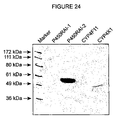

Classifications
-
- G—PHYSICS
- G01—MEASURING; TESTING
- G01N—INVESTIGATING OR ANALYSING MATERIALS BY DETERMINING THEIR CHEMICAL OR PHYSICAL PROPERTIES
- G01N33/00—Investigating or analysing materials by specific methods not covered by groups G01N1/00 - G01N31/00
- G01N33/48—Biological material, e.g. blood, urine; Haemocytometers
- G01N33/50—Chemical analysis of biological material, e.g. blood, urine; Testing involving biospecific ligand binding methods; Immunological testing
- G01N33/53—Immunoassay; Biospecific binding assay; Materials therefor
- G01N33/573—Immunoassay; Biospecific binding assay; Materials therefor for enzymes or isoenzymes
-
- A—HUMAN NECESSITIES
- A61—MEDICAL OR VETERINARY SCIENCE; HYGIENE
- A61P—SPECIFIC THERAPEUTIC ACTIVITY OF CHEMICAL COMPOUNDS OR MEDICINAL PREPARATIONS
- A61P35/00—Antineoplastic agents
-
- C—CHEMISTRY; METALLURGY
- C07—ORGANIC CHEMISTRY
- C07K—PEPTIDES
- C07K16/00—Immunoglobulins [IGs], e.g. monoclonal or polyclonal antibodies
- C07K16/18—Immunoglobulins [IGs], e.g. monoclonal or polyclonal antibodies against material from animals or humans
- C07K16/28—Immunoglobulins [IGs], e.g. monoclonal or polyclonal antibodies against material from animals or humans against receptors, cell surface antigens or cell surface determinants
- C07K16/30—Immunoglobulins [IGs], e.g. monoclonal or polyclonal antibodies against material from animals or humans against receptors, cell surface antigens or cell surface determinants from tumour cells
-
- C—CHEMISTRY; METALLURGY
- C07—ORGANIC CHEMISTRY
- C07K—PEPTIDES
- C07K16/00—Immunoglobulins [IGs], e.g. monoclonal or polyclonal antibodies
- C07K16/40—Immunoglobulins [IGs], e.g. monoclonal or polyclonal antibodies against enzymes
-
- C—CHEMISTRY; METALLURGY
- C12—BIOCHEMISTRY; BEER; SPIRITS; WINE; VINEGAR; MICROBIOLOGY; ENZYMOLOGY; MUTATION OR GENETIC ENGINEERING
- C12N—MICROORGANISMS OR ENZYMES; COMPOSITIONS THEREOF; PROPAGATING, PRESERVING, OR MAINTAINING MICROORGANISMS; MUTATION OR GENETIC ENGINEERING; CULTURE MEDIA
- C12N9/00—Enzymes; Proenzymes; Compositions thereof; Processes for preparing, activating, inhibiting, separating or purifying enzymes
- C12N9/0004—Oxidoreductases (1.)
- C12N9/0071—Oxidoreductases (1.) acting on paired donors with incorporation of molecular oxygen (1.14)
- C12N9/0077—Oxidoreductases (1.) acting on paired donors with incorporation of molecular oxygen (1.14) with a reduced iron-sulfur protein as one donor (1.14.15)
-
- G—PHYSICS
- G01—MEASURING; TESTING
- G01N—INVESTIGATING OR ANALYSING MATERIALS BY DETERMINING THEIR CHEMICAL OR PHYSICAL PROPERTIES
- G01N33/00—Investigating or analysing materials by specific methods not covered by groups G01N1/00 - G01N31/00
- G01N33/48—Biological material, e.g. blood, urine; Haemocytometers
- G01N33/50—Chemical analysis of biological material, e.g. blood, urine; Testing involving biospecific ligand binding methods; Immunological testing
- G01N33/53—Immunoassay; Biospecific binding assay; Materials therefor
- G01N33/574—Immunoassay; Biospecific binding assay; Materials therefor for cancer
- G01N33/57407—Specifically defined cancers
- G01N33/57423—Specifically defined cancers of lung
-
- A—HUMAN NECESSITIES
- A01—AGRICULTURE; FORESTRY; ANIMAL HUSBANDRY; HUNTING; TRAPPING; FISHING
- A01K—ANIMAL HUSBANDRY; CARE OF BIRDS, FISHES, INSECTS; FISHING; REARING OR BREEDING ANIMALS, NOT OTHERWISE PROVIDED FOR; NEW BREEDS OF ANIMALS
- A01K2217/00—Genetically modified animals
- A01K2217/05—Animals comprising random inserted nucleic acids (transgenic)
-
- G—PHYSICS
- G01—MEASURING; TESTING
- G01N—INVESTIGATING OR ANALYSING MATERIALS BY DETERMINING THEIR CHEMICAL OR PHYSICAL PROPERTIES
- G01N2800/00—Detection or diagnosis of diseases
- G01N2800/52—Predicting or monitoring the response to treatment, e.g. for selection of therapy based on assay results in personalised medicine; Prognosis
-
- Y—GENERAL TAGGING OF NEW TECHNOLOGICAL DEVELOPMENTS; GENERAL TAGGING OF CROSS-SECTIONAL TECHNOLOGIES SPANNING OVER SEVERAL SECTIONS OF THE IPC; TECHNICAL SUBJECTS COVERED BY FORMER USPC CROSS-REFERENCE ART COLLECTIONS [XRACs] AND DIGESTS
- Y10—TECHNICAL SUBJECTS COVERED BY FORMER USPC
- Y10S—TECHNICAL SUBJECTS COVERED BY FORMER USPC CROSS-REFERENCE ART COLLECTIONS [XRACs] AND DIGESTS
- Y10S435/00—Chemistry: molecular biology and microbiology
- Y10S435/81—Packaged device or kit
Definitions
- the present invention relates to a novel gene encoding a polypeptide that is a member of the cytochrome P450 family. More particularly the present invention relates to a polynucleotide encoding the novel polypeptide, to an antisense polynucleotide thereof and to fragments thereof. The invention further relates to the novel cytochrome P450 as well as to vectors, host cells and antibodies to the polypeptide and the recombinant methods for producing the same. Uses and methods relating to any of the foregoing are also included within the scope of the invention.
- the cytochromes P450 comprise a large gene superfamily that encodes over 500 distinct heme-thiolate proteins that catalyze the oxidation of drugs and numerous other compounds in the body [Nelson et al., (1996); Guengerich (1991)]. Since there are at least 500 different cytochrome P450 enzymes, it is of considerable interest in the pharmaceutical and other fields to identify which of these enzymes are most important in the metabolism of individual compounds. There are now numerous examples of adverse drug-drug interactions and side effects that can now be understood in terms of the cytochrome P450 enzymes.
- P450 proteins are ubiquitous in living organisms, and have been identified in bacteria, yeast, plants and animals [Nelson et al (1996); and Nelson, (1999a)].
- the P450 enzymes catalyze the metabolism of a wide variety of drugs, xenobiotics carcinogens, mutagens, and pesticides, and are responsible for the bioactivation of numerous endogenous compounds including steroids, prostaglandins, bile acids and fatty acids body [Nelson et al., (1996); Guengerich (1991); Nebert et al., (1989)].
- Cytochrome P450 metabolism of xenobiotics can result in detoxification of toxic compounds by their conjugation into excretable forms or can result in activation of compounds into metabolites that are toxic, mutagenic, or carcinogenic. Many steroids are deactivated by cytochrome P450-catalyzed oxidation.
- Vitamin A metabolism gives rise to several active forms of retinoic acid (RA) which are involved in regulating gene expression during development, regeneration, and in the growth and differentiation of epithelial tissues.
- RA retinoic acid
- RA itself has been found to be useful therapeutically, notably in the treatment of cancers, including acute promyelocytic leukemia (APL), tumors of the head and neck, and skin cancer, as well as in the treatment of skin disorders such as the premalignancy associated actinic keratoses, acne, psoriasis and ichthyosis.
- APL acute promyelocytic leukemia
- RA increases the rate of its own metabolism which in turn results in the inability to sustain effective therapeutic doses of RA.
- Therapeutic administration of RA can result in a variety of undesirable side effects and it is therefore important to establish and maintain the minimal requisite doses of RA in treatment.
- RA treatments during pregnancy can lead to severe teratogenic effects on the fetus.
- Adverse reactions to RA treatment also include headache, nausea, chelitis, facial dermatitis, conjunctivitis, and dryness of nasal mucosa.
- Prolonged exposure to RA can cause major elevations in serum triglycerides and can lead to severe abnormalities of liver function, including hepatomegaly, cirrhosis and portal hypertension.
- RA metabolism may also account for the lack of response of certain tumors to RA treatment.
- cytochrome P450 inhibitors that block RA metabolism, resulting in increased tissue levels of RA, may be useful therapeutic agents in the treatment of prostate cancer [Wouters, 1992; De Coster, 1996].
- RA metabolizing cytochrome P450s may be useful targets for the treatment of a number of different types of cancer.
- the catabolism of RA is thought to be initiated by hydroxylation either at the C4-, or C18-position of the ⁇ -ionone ring of RA [Napoli, 1996].
- the C4-hydroxylation step is mediated by cytochrome P450 activity, as judged by the ability of broad spectrum P450 inhibitors such as ketoconazole and liarazole to block 4-hydroxylation [Williams, 1987, Van Wauwe, 1988; Van Wauwe, 1990, Van Wauwe, 1992, Wouters, 1992].
- RA metabolism can be induced by RA pretreatment [Frolik, 1979, Roberts, 1979a and b; Duell, 1992; Wouters, 1992].
- Studies involving targeted disruption of RAR genes in F9 cells suggest that RAR- ⁇ and RAR- ⁇ isoforms may play a role in regulating the enzymes responsible for this increased metabolism [Boylan, 1995].
- 4-oxoretinol (4-oxo-ROL) can have greater biological activity than retinol.
- the 4-oxo-ROL is inducible by RA in F9 and P19 mouse teratocarcinoma cells [Blumberg et al., 1995; Achkar et al., 1996].
- retinoic acid retinoic acid
- cytochrome P450 enzyme Several P450s have since been shown to metabolize RA, including P450 proteins from human, zebrafish and mouse.
- human P450RAI which is induced by RA, metabolizes RA to more poplar derivatives including 4-hydroxy retinoic acid (4-OHRA) and 4-oxo retinoic acid (4-oxo RA) [White et al. (1996a)]. Since RA is useful as an antitumor agent, it is desirable to maintain high tissue levels of RA.
- cytochrome P450 inhibitors that block RA metabolism, resulting in increased tissue levels of RA may be useful therapeutic agents in the treatment of cancers, such as prostate cancer [Wouters et al., (1992); and De Coster et al., (1996)].
- P450RAI gene is an important step in the understanding of RA signaling but its presence has been known since Roberts et al. (1979a) first postulated that the catabolism of RA was mediated by a P450 enzyme [Frolik et al., 1979; Roberts et al., 1979a]. More recently, the isolation of cDNAs which encode the full-length human and mouse P450RAI orthologs whose expression, like that of the fish cytochrome, is highly inducible by RA has been achieved [Fujii et al., 1997; Ray et al., 1997]. Human and mouse genomic P450RAI-1 sequences are identified herein as SEQ ID NOS: 15 and 16.
- the mouse sequence encoding P450RAI-1 is identified herein as SEQ ID NO: 17. Homologs have also been isolated from human, mouse, chick and xenopus all exhibiting a high degree of sequence conservation [Abu-Abed et al., 1998; Hollermann et al., 1998; White et al., 1997]. There is extensive identity between the human and fish P450RAI genes which overall is 68% at the amino acid level (over 90% between mouse and human).
- MCF7 cells have been shown to have RA inducible RA metabolism [Butler and Fontana, 1992; Wouters et al., 1992].
- the expression of P450RAI in these cells is dependent on the continuous presence of RA [White et al., 1997]. This suggests that P450RAI regulation by RA forms an autoregulatory feedback loop that functions to limit local concentrations of RA, such that when normal physiological levels of RA are exceeded, induction of P450RAI acts to normalize RA levels.
- the inducible expression of P450RAI in mouse embryos also suggests that a similar autoregulatory mechanism may limit exposure to RA sensitive tissues during development [Iulianella et al., 1999].
- All-trans-RA is a critical regulator of gene expression during embryonic development and in the maintenance of adult epithelial tissues [Gudas, et al. (1994).; Lotan, R. M. (1995); Lotan, R. (1996); Morriss-Kay, G. M. (1996)).
- the effects of all-trans-RA are mediated by heterodimers of nuclear receptors for retinoic acid (RARs) and retinoid-X-receptors, which are regulated by by the 9-cis isomer of RA.
- RAR ⁇ , ⁇ and ⁇ Three different subtypes exist for each of these receptors (RAR ⁇ , ⁇ and ⁇ ; RXR RAR ⁇ , ⁇ and ⁇ ) which individually are expressed in a tissue specific manner but collectively can be found in essentially all cell types, both during embryonic development and in the adult [Chambon, P. (1995).].
- the activity of RA in these tissues is controlled, to a large extent, by enzymes involved in its synthesis from retinaldehyde (ALDH-1 and RAIDH-2) and its catabolism to 4-OH, 4-oxo and 18-OH products (P450RAI) [White J. A., et al. (1997); connella, A. et al. (1999); McCaffery P. et al., (1999) Niederreither, K. et al. (1999) Swindell E., et al. (1999)].
- P450RAI-1 (CYP26A) from zebrafish, mouse, human, chick and xenopus which is responsible for the metabolism of active all-trans-RA to inactive polar metabolites including 4-OH-RA, 4-oxo-RA and 18-OH-RA [White J., et al. (1997); Swindell E., et al. (1999); White, J. & Petkovich, M. (1996); Abu-Abed, et al.(1998); Fujii, H. et al., (1997); Ray, W. et al. (1997); Hollermann, T et al. (1998)].
- P450RAI-1 expression can be induced by all-trans-RA pre-treatment in multiple tissues, and cell types, and this expression is concomitant with increased all-trans-RA catabolism.
- MCF7 cells all all-trans-RA suggesting a feedback-loop mechanism is dependent on the continued presence of all-trans-RA suggesting a feedback-loop mechanism for the regulation of all-trans-RA levels [White J., et al. (1997)].
- Inducible expression of P450RAI-1 has also been observed in vivo in zebrafish, chick, xenopus and mouse embryos suggesting that this autoregulatory feedback-loop plays an important role in balancing all-trans-RA levels in certain developing tissues.
- RALDH-2 expressing tissues have been shown to contain retinoid activity as measured by both retinoid responsive reporter gene activity and direct measurement of RA levels from tissue extracts; by similar analyses, P450RAI-1 expressing tissues do not [Iulianella, A et al. (1999); McCaffery P. et al. (1999)].
- P450RAI-1 expressing tissues do not [Iulianella, A et al. (1999); McCaffery P. et al. (1999)].
- over expression of P450RAI-1 in xenopus embryos has been shown to abrogate the teratogenic effects of exogenously applied RA, consistent with a catabolic role for its enzyme [Hollermann, T et al. (1998)].
- the present inventors have identified and characterized a novel cytochrome P450 that can metabolize retinoic acid, preferably all-trans retinoic acid, (hereinafter referred to as P450RAI-2) and the nucleic acid sequence encoding therefor.
- P450RAI-2 all-trans retinoic acid
- the novel cytochrome P450 metabolizes all-trans retinoic acid from into more polar metabolites.
- the P450RAI-2 is a mammalian P450RAI-2, and preferably a murine (such as comprising amino acid sequence depicted in SEQ. ID. NO. 11 or 17), rat (such as that comprising SEQ. ID. NO. 27) or human P450RAI-2(such as that of SEQ. ID. NO. 5).
- the P450RAI-2 is a zebrafish P450RAI-2 (such as that comprising SEQ. ID. NO. 12).
- the invention provides functional homologs of the above-noted sequences, or functional homologous fragments thereof.
- the P450RAI-2 and encoding nucleic acid sequence of the invention can be isolated and characterized from a number of different tissues such as spleen, kidney, skeletal muscle, brain, liver, retina, heart or small intestine, it is preferably isolated and characterized from brain cells, such as from from the cerebellum, cerebal cortex, medulla, occipital pole, frontal lobe and temporal lobe, and most preferably from the cerebellum.
- P450RAI-2 is isolated from lung cells, preferably tumour lung cells, such as human lung tissue or SKMES cells.
- the present invention provides an isolated nucleic acid molecule comprising a sequence encoding a P450RAI-2, preferably a human P450RAI-2 (such as SEQ. ID. NO. 4 or 28 or degenerate or corresponding RNA sequences (where T is U) thereof) or a murine P450RAI-2 (such as comprising any one of SEQ. ID. NO. 17–26 or degenerate or corresponding RNA sequences (where T is U)) or fragments thereof, preferably fragments encoding functional fagments of P450RAI-2.
- a human P450RAI-2 such as SEQ. ID. NO. 4 or 28 or degenerate or corresponding RNA sequences (where T is U) thereof
- a murine P450RAI-2 such as comprising any one of SEQ. ID. NO. 17–26 or degenerate or corresponding RNA sequences (where T is U)
- fragments thereof preferably fragments encoding functional fagments of P450RAI-2
- an isolated nucleic acid molecule having: (a) a nucleic acid sequence as shown in SEQ ID NOS: 4, 5, 28, 27, 7, 18, 26, 21, 22, 23, 24, or 25 where T can also be U; (b) nucleic acid sequences complementary to (a); (c) nucleic acid sequences which are homologous to (a) and (b); (d) a nucleic acid molecule differing from any of the nucleic acid molecules of (a) to (c) in codon sequences due to the degeneracy of the genetic code; or (e) a fragment of (a) to (d) that is at least 15 bases, preferably 20 to 30 bases, and which will hybridize to (a) to (c) under stringent hybridization conditions.
- the present invention also includes the P450RAI-2 polypeptide itself. Accordingly, the invention provides a polypeptide having an amino acid sequence of a P450RAI-2. Preferably, the invention provides a polypeptide having either the human (SEQ ID NO: 5) or a homologous mouse sequence or fragment thereof. The invention also comprises peptides comprising fragments of the amino acid sequence of SEQ ID NO: 5. Preferably the fragments comprise amino acid sequences of SEQ ID NOS: 11 or 12 or encoded by nucleic acid sequences SEQ ID NOS. 18, 26, or 21 to 25. In another embodiment the fragments preferably comprise at least 14 amino acid residues and are most preferably antigenic or immunogenic.
- the invention provides peptides encoded by a nucleic acid sequence of SEQ ID NO: 4 or fragments thereof and to an antisense nucleic acid molecule to all or part of the nucleic acid molecule encoding P450RAI-2.
- homologs, analogs, and modifed proteins of the invention as described herein are encompassed within the scope of this invention.
- the invention also provides a nucleic acid molecule of the invention operationally linked to an expression control sequence in a suitable expression vector.
- the expression vector comprising the nucleic acid molecule of the invention is capable of being activated to express the peptide which is encoded by the nucleic acid molecule and is capable of being transformed or transfected into a suitable host cell. Such transformed or transfected cells are also encompassed with the scope of this invention.
- the invention also provides a method of preparing a P450RAI-2 protein of the invention utilizing a nucleic acid molecule of the invention.
- a method for preparing a P450RAI-2 protein of the invention comprising: transforming a host cell with a recombinant expression vector comprising a nucleic acid sequence of the invention; (b) selecting transformed host cells from untransformed host cells; (c) culturing a selected transformed host cell under conditions which allow expression of the protein; and (d) isolating the protein.
- the invention also encompasses an antibody specific for one or more epitopes of a protein of the invention, such as a peptide specific antibody or a polyclonal antibody, and more preferably a monoclonal antibody.
- the invention also encompasses methods for preparing the antibodies.
- the epitopes are selected from the group consisting of SEQ ID NOS: 5, 11, 12, or those encoded by nucelic acid sequences SEQ ID NOS; 4, 6, 8, 18, 21–25, 27 or 28 or immunogenic fragments thereof.
- the invention also includes a method for detecting a disease or medical condition associated with P450RAI-2 expression or RA metabolism in an animal.
- a disease or medical condition associated with P450RAI-2 expression means any disease that can be affected or characterized by the level of P450RAI-2 expression. This includes, without limitation, diseases affected by, high, normal, reduced or non-existent expression of P450RAI-2 or expression of mutated P450RAI-2.
- a disease or medical condition associated with P450RAI-2 expression includes diseases associated with RA metabolism such as cell cycle regulation, particularly cell growth and apoptosis, for instance cancer, dysplasia, autoimmune disease, dermatological disorders and disabilities associated with high order brain functions, such as learning and memory.
- the method comprises assaying for the P450RAI-2 from a sample, such as a blood sample, a biopsy, or other cellular or tissue sample, from an animal susceptible of having such a disease.
- the method comprises contacting the sample with an antibody of the invention that binds P450RAI-2, and measuring the amount of antibody bound to P450RAI-2 in the sample, or unreacted antibody.
- the method involves detecting the presence of a nucleic acid molecule having a sequence encoding a P450RAI-2, comprising contacting the sample with a nucleotide probe which hybridizes with the nucleic acid molecule, preferably mRNA or cDNA to form a hybridization product under conditions which permit the formation of the hybridization product, and assaying for the hybridization product.
- the invention further includes a kit for detecting a disease or condition associated with P450RAI-2 expression in a sample comprising an antibody of the invention, preferably a monoclonal antibody. Preferably directions for its use is also provided.
- the kit may also contain reagents that are required for binding of the antibody to a P450RAI-2 protein in the sample.
- the invention also provides a kit for detecting the presence of a nucleic acid molecule having a sequence encoding a polypeptide related to or analogous to a polypeptide of the invention, comprising a nucleotide probe which hybridizes with the nucleic acid molecule, reagents required for hybridization of the nucleotide probe with the nucleic acid molecule, and directions for its use.
- the invention further provides a method of treating or preventing a medical condition disease associated with P450RAI-2 or RA expression comprising administering an effective amount of an agent that activates, simulates or inhibits P450RAI-2 expression, as the situation requires, to an animal in need thereof.
- P450RAI-2, a therapeutically active fragment thereof, or an agent which activates or simulates P450RAI-2 expression is administered to the animal in need thereof to treat cancer, dysplasia, autoimmune disease or dermatological disorders or to improve high order brain functions or dermatological disorders that is related to the presence of too much RA or all-trans-RA.
- the disease is associated with over expression of P450RAI-2 or too little RA, or all-trans-RA or apoptosis
- the method of treatment comprises adminstration of an effective amount of an agent that inhibits P450RAI-2 expression such as an antibody to P450RAI-2, a mutation thereof, or an antisense nucleic acid molecule to all or part of the P450RAI-2 gene.
- the invention provides pharmaceutical compositions comprising the nucleic acid molecules of proteins, antibodies, vectors or cells of the invention and a pharmaceutically acceptable carrier.
- the invention further provides a method for identifying modulators of P450RAI-2 expression or P450RAI-2 activity.
- this involved incubating the gene under conditions conducive to gene expression with a potential modulator of gene expression and then assaying, for instance by southern, norther or western blot for the presence of expression of the gene and the effect of the modulator on such expression (increased expression, inhibited expression or no effect on expression).
- the effect of the modulator on such expression is determined by comparison to various controls run without the potential modulator and/or with the potential modulator but without the gene.
- Expression of P450RAI-2 can also be done by radioactive or othe labeling for such expression or by antibody screening, such as using the P450RAI-2 polyclonal antibody discloses herein.
- the invention further provides a method of rational drug design.
- the invention provides methods or uses of the proteins and nucleic acid molecules of the invention, such as in relation to medical treatment (where inhibitors of P450RAI-2, such as antibodies or antisense molecules can be used to treat a condition associated with too much RA metabolism (e.g., too little RA), preferably all-trans RA metabolism.
- P450RAI-2 gene or protein or stimulators or functional fragments or homologs thereof can be used in cases where there is too much RA or all-trans-RA.
- FIG. 1 is a schematic diagram showing the 6 exons of human cytochrome p450RAI-2 on human clone having GenBank Accession No. AC007002 (SEQ ID NO:3).
- the numbers above the schematic diagram indicate the amino acid regions of the exons and the numbers below the schematic refer to nucleotide positions on the human sequence.
- FIG. 2 Human P450RAI-2 cDNA and amino acid sequence (SEQ ID NO: 28) comprising the coding sequence and the 3′ untranslated sequence.
- the full-length cDNA clone was isolated from human retina cDNA.
- the deduced primary sequence using single-letter amino acid codes, identifies a 512 amino acid protein which is shown directly below the corresponding nucleotide sequence. Numbers on the right refer to the nucleotide positions.
- FIG. 3 Amino acid eequence comparison between human P450RAI-2 (SEQ. ID. NO. 5) and human P450RAI-1 (amino acid sequence shown in SEQ. ID. NO. 13). Predicted amino acid sequences were aligned using Omiga software (Oxford Molecular, CA). Identical amino acids are highlighted by the black boxes with white letters. Conservatively substituted amino acids are indicated by the open boxes. Gaps, indicated by the dashes, have been introduced in several regions in order to optimize amino acid identity between the two proteins. Overall the two protein sequences show 42% identity at the amino acid level. Numbers on the right hand side refer to the corresponding amino acid positions.
- FIG. 4 Expression of P450RAI-2 in human tissues.
- RNA samples from 76 normal human tissues were probed for expression of P450RAI-2 transcripts using a commercially available dot blot (A). Signals representing P450RAI-2 transcripts are observed in three samples, a2, b2 and h1 representing left and right cerebellum and pons respectively.
- Sample a1 is whole brain and a11 is fetal brain for comparison.
- Control hybridization with a human ubiquitin probe (B) shows differences in RNA loading.
- Northern blot analyses for expression of P450RAI-2 expression in human brain D). 2 ⁇ g poly A+ mRNA each from eight different human brain tissues was used to analyze P450RAI-2 expression. Transcripts at approximately 5 Kb corresponding to P450RAI-2 are indicated, with the highest levels of expression seen in cerebellum.
- FIG. 4C shows the location of the different human mRNAs on the blot used in FIG. 4A .
- FIG. 5 shows northern blot analysis of human cell lines probed with an ⁇ -[ 32 P]-dATP labeled probe having the sequence identified as SEQ ID NO:4 SKMES; CALU-1 and MCF-7 (A).
- Cells were treated with DMSO, all-trans retinoic acid, 9-cis retinoic acid or 13-cis retinoic acid.
- Cells were exposed to 10 ⁇ 6 M final concentrations of each retinoid dissolved in DMSO.
- Transcripts corresponding to P450RAI-2 are indicated by the arrowhead.
- the blot was also probed with the human ⁇ -actin probe to control for RNA loading of the gel, shown in the middle panel (B).
- the 18S and 28S portions of the mRNA as seen on the ethidium bromide stained agarose gel are shown in the bottom panel (C).
- FIG. 6 is similar to FIG. 5 except that the cells, HPK1A-RAS, were treated with either DMSO or all-trans retinoic acid.
- FIG. 7 All-trans-RA induction of P450RAI-2 expression.
- P450RAI-2 hybridizing transcripts are identified.
- the positions of 28S and 18S ribosomal RNA are indicated along with the ethidium bromide stained gel showing the relative abundance of RNA in all samples.
- Multiple other cell lines were tested using semiquantitative RT-PCR and southern blotting (B). Transcripts for P450RAI-2 are regulated in a cell specific manner.
- P450RAI-2 is constitutively expressed in SK-MES-1 and SW900 and possibly WTE; inducible in MCF-7, HPK1a-ras and HeLa; and undetectable in SK-Luci-6, V79 or NB4.
- FIG. 8A shows the total aqueous soluble radioactivity measured using aliquots of the aqueous soluble extracts from media alone, as well as cells transfected with pcDNA3.1, pcDNA3.1-CYP26A (human) (i.e., P450RAI) or pcDNA-P450RAI-2.
- Aqueous soluble extracts were subjected to ⁇ -scintillation counting.
- FIG. 8B shows a summary of the peak integration analysis performed after HPLC on the organic soluble radioactivity. Samples were the same as in FIG. 8A .
- Cells transfected with either pcDNA3.1-CYP26A (human) or pcDNA3.1-P450RAI-2 show a decrease in all-trans retinoic acid substrate with a concomitant increase in the production of 4-OH-retinoic acid, 4-oxo-retinoic acid and more polar peaks.
- FIG. 9 shows the same analysis as FIG. 8B except that cells were treated with 1 ⁇ M non-radioactive all-trans-retinoic acid.
- FIG. 10 shows a control HPLC trace from media alone in the absence of the COS-1 cells.
- An all-trans-retinoic acid substrate peak is observed at 20.808 minutes. Peaks at 1.409 and 1.874 are contaminants not characteristic of retinoids.
- the top panel shows photo diode array detection of the peaks. Retinoids have characteristic UV maxima in the range of about 320 to about 380 nM.
- FIG. 11 is similar to FIG. 10 except that the sample is from COS-1 cells transfected with the pcDNA3.1 plasmid alone.
- FIG. 12 is similar to FIG. 10 except that the sample is from COS-1 cells transfected with pcDNA3.1-P450RAI. Multiple more polar peaks characteristic of retinoids are generated in these cells. Peaks which co-elute with standards for 4-OH-retinoic acid and 4-oxo-retinoic acid were observed at 8.191 and 9.661 minutes respectively. Additional peaks which have not been characterized are also evident.
- FIG. 13 is similar to FIG. 12 except that the sample is from COS-1 cells transfected with pcDNA3.1-P450RAI-2.
- FIG. 14 shows HPLC analysis of all-trans-RA metabolism.
- COS-1 cells were transiently transfected with pcDNA3.1-P450RAI-2 (A), pcDNA3.1-P450RAI-1 (B) or pcDNA3.1 alone (C) and exposed to ⁇ M all-trans-RA for 3 hours. Incubation with RA was followed by total lipid extraction of the media and subsequent HPLC analysis using a reverse-phase system. Expression of either P450RAI-2 or P450RAI-1 in cells causes disappearance of all-trans-RA substrate (compare C to A or B) in addition to the generation of more polar metabolic products. Identities of the retinoids.
- Peaks labeled 1–4 (A) have spectral properties characteristic of retinoids, specifically a UV maxima of between 320 and 350 nm. The exact identity of these polar metabolites remains to be established.
- FIG. 15 shows metabolism of all-trans-RA.
- COS-1 cells transiently transfected with pcDNA3.1 -P450RAI-2, pcDNA3.1-P450RAI-1 or pcDNA3.1 alone and exposed to 100 nM [ 3 H]all-trans-RA for 3 hours.
- Conversion of all-trans-RA to aqueous-soluble metabolites is observed in P450RAI-2 or P450RAI-1 expressing cells compared to pcDNA vector or media alone control samples (A).
- HPLC analysis with subsequent ⁇ -scintillation counting was used to evaluate the lipid-soluble retinoids generated from P450RAI-2 or P450RAI-1 expressing cells.
- FIGS. 16 A–E is competitive inhibition of P450RAI-mediated all-trans-RA metabolism.
- COS 1 cells transiently transfected with pcDNA3.1-P450RAI-2, pcDNA3.1-P450RAI-1 or pcDNA3.1 alone were used to assess the ability of several retinoids to competitively inhibit all-trans-RA metabolism.
- Cells were exposed to 2 nM [ 3 H]all-trans-RA with increasing concentrations of unlabelled competitor and assayed for generation of aqueous soluble radioactivity.
- Panels C and D show cells expressing P450RAI-2 with panels A and B show cells expressing P450RAI-1.9-cis-RA and 13-cis-RA (top panels 16 A and 16 C) are less effective than all-trans-RA as competitors for either P450RAI-2 or P450RAI-1.
- FIG. 16E shows [ 3 ]-RA-metabolism inhibition assay using microsomes prepared from Hela-CYP26B(stable) cells. Microsomes (5 micograms) were incubated with [ 3 H]-RA and increased concentration o funlabeled all-trans-RA, 9-cis-RA, 13-cis-RA and ketoconazole. Retinol and retinal were similarly tested (bottom panels 16 B and 16 D) and found to be less effective competitors. For comparison, the nonspecific cytochrome P450 inhibitor ketoconazole is shown in each panel.
- FIG. 17 shows Northern blot (A) and RT-PCR (B) results for total RNA samples from the various cell samples designated.
- FIG. 18 shows expression of P450RAI-2 in 8.0 and 8.5 dpc mouse embryos.
- A 8.0 dpc lateral view. No apparent staining.
- B 8.0 dpc, dorsal view. Staining at anterior end of neutral folds.
- C 8.5 dpc, lateral view. Expression is evident, possibly in presumptive rhombmeres 2, 5, and 6.
- D 8.5 dpc, dorsal view, rhombomere expression of RAI2 is clearly evident.
- TL Tail; YS Yolk Sac; NF: Folds; pr: Presumptive Rhombomeres.
- FIG. 19 shows expression of P450RAI2 in 9.0 and 10.5 dpc mouse embyros.
- A 9.0 dpc, lateral view. Specific staining is visible in the eye, and rhombomeres 5 and 6. Diffuse staining is visible where the hind bud is beginning to form.
- B 9.0 dpc, dorsal view. Rhombomeres 5 and 6 show RAI2 expression.
- C 10.5 dpc, lateral view. The otic vesicle and eye are stained.
- D 10.5 dpc, dorsal view. Specific staining is observed in both otic vesicles as well as as the hind linb bud.
- HL hind limb bud
- OV Otic Vesicle.
- FIG. 20 shows expression of P450RAI2 staining in 11.5 dpc mouse embryo(A) 11.5 dpc, laterial view, staining is visible in both the fore and hindlimb bud.
- FIG. 21 shows P450RAI2 staining in embryos treated with Retinoic Acid
- A 8.5 dpc, lateral view, staining is observed in rhombomere 5 and the tail mesoderm as indicated by the arrow.
- B 8.5 dpc, dorsal view clearly showing P450RAI2 expression in rhombomere 5.
- C 9.5 dpc, lateral view, expresion of P450RAI2 is observed in rhombomeres 5 and 6, the developing hindlinb, somites and posterior mesoderm.
- FIG. 22 shows P450RAI2 expression in 11.5 dpc embryos treated with retinoic acid.
- A 11.5 dpc, lateral view, P450RAI2 expression is observed in both the developing hind and fore limb.
- B 11.5 dpc, ventral view, as in embryos untreated with retinoic acid, P450RAI2 expression is not observed in the aptical ectodermal ridge.
- FIG. 23 shows P450RAI2 (CYP26B), P450RAI-1(CYP26A) and P450RAI-3 (CYP26C) expression in normal (N) and tumour (T) lung tissue.
- B-actin is a control.
- FIG. 24 is an immunoblot indicating P450RAI-2 specificity of the polyclonal antibody made against CYP26B (P450RAI-2) as compared to other cytochromes (CYP26A, CYP4F11, and CYP4X1).
- FIG. 25 shows the RT-PCR assay of CYP26A (P450RAI-1) and CYP26B (P450RAI-2) expression in human retina and MCF-7 cells.
- Retinoids particularly all-trans retinoic acid (all-trans-RA) are potent regulators of cell differentiation, cell proliferation and apoptosis.
- all-trans-RA all-trans retinoic acid
- the control of all-trans-RA in cells and tissues is regulated by the balance between its biosynthesis and its catabolism to inactive metabolites.
- the cytochrome P450 enzyme P450RAI-1 is partially responsible for this inactivation of all-trans-RA.
- P450RAI-2 a second related enzyme, here termed P450RAI-2, which is also involved in the specific inactivation or metabolism of all-trans-RA.
- a cDNA has been isolated from human retina and sequenced.
- a protein encoded by the cDNA has been expressed and shown to have the ability to metabolize all-trans-RA to more polar metabolites.
- the metabolites so formed include products that have been oxidized at the 4-position of the ⁇ -ionone ring, including the corresponding acid hydroxylated at the 4-position of the ⁇ -ionone ring.
- the MRNA has been found to be inducible in multiple cell types including but not limited to human lung and human skin upon the addition of all-trans retinoic acid. It has also been shown to be expressed in human brain tissue and breast and lung cancer cells.
- the present inventors herein show that tansiently transfected P450RAI-2 can convert all-trans-RA to more polar metabolites including 4-oxo, 4-OH, and 18-OH-retinoic acid.
- Competition experiments with other retinoids suggest that all-trans-RA is the preferred substrate, but not necessarily the only retinoid substrate.
- retinoids as used herein means a group of compounds which includes retinoic acid, vitamin A (retinol) and a series of natural and synthetic derivatives that can exert profound effects on development and differentiation in a wide variety of systems.
- retinoid is also intended to encompass an equivalent thereof having the same functional characteristics which may be produced, for example, by computational chemistry.
- A Ala—alanine
- C Cys—cysteine
- D Asp—aspartic acid
- E Glu—glutamic acid
- F Phe—phenylalanine
- G Gly—glycine
- H His—histidine
- I Ile—isoleucine
- K Lys—lysine
- L Leu—leucine
- M Met—methionine
- N Asn—asparagine
- Q Gln—glutamine
- R Arg—arginine
- S Ser—serine
- T Thr—threonine
- V Val—valine
- W Trp—tryptophan
- Y Tyr—tyrosine
- p.Y. P.Tyr—phosphotyrosine.
- the present invention provides an isolated nucleic aid molecule comprising a sequence encoding a cytochrome P450RAI-2 polypeptide.
- isolated refers to a nucleic acid substantially free of cellular material or culture medium when produced by recombinant DNA techniques, or chemical precursors, or other chemicals when chemically synthesized.
- nucleic acid molecule is intended to include unmodified DNA or RNA or modified DNA or RNA.
- the nucleic acid molecules or polynucleotides of the invention can be composed of single- and double stranded DNA, DNA that is a mixture of single- and double-stranded regions, single- and double stranded RNA, and RNA that is a mixture of single- and double-stranded regions, hybrid molecules comprising DNA and RNA that may be single-stranded or, more typically double stranded or a mixture of single- and double stranded regions.
- the P450RAI-2 nucleic acid molecules can be composed of triple-stranded regions comprising RNA or DNA or both RNA and DNA.
- the nucleic acid molecules of the invention may also contain one or more modified bases or DNA or RNA backbones modified for stability or for other reasons.
- “Modified” bases include, for example, tritiated bases and unusual bases such as inosine.
- a variety of modifications can be made to DNA and RNA; thus “nucleic acid molecule” embraces chemically, enzymatically, or metabolically modified forms.
- polynucleotide shall have a corresponding meaning.
- nucleic acid molecule comprising a nucleotide sequence selected from the group consisting of:
- a fragment of (a) to (d) that is at least 15 bases, preferably 20 to 30 bases, and which would hybridize to (a) to (d) under stringent hybridization conditions.
- the fragment is at least 35, 40, 45, 50, 55, or 60 or more nucelotides in length.
- the fragment is capable of oxidizing a retinoid.
- it preferably has at least about 55 percent homology with the protein encoded by a nucleotide sequence of (a) or (b).
- the nucleotide sequence in (a) above is a human P450RAI-2 sequence.
- the nucleic acid sequence can be used as a probe or a primer, such as for PCR, like SEQ ID NOS: 9, 10, 29, 30, 33, 37 or 38, more preferably SEQ. ID. Nos. 37 and/or 38.
- the nucleotide sequence of (a) above is selected from the group consisting of: a mouse P450RAI-2 sequence, such as SEQ ID NO: 6, 18, 26, 21, 22, 23, 24 or 25; a rat P450RAI-2 sequence, SEQ ID NO: 27; or a zebrafish P450RAI-2 sequence, SEQ ID NO: 8.
- the nucleic acid molecule of the invention encodes a protein that is capable of oxidizing a retinoid, preferably retinoic acid, preferably at the 4-position of the ⁇ -ionone ring.
- the nucleic acid molecule of the invention encodes a protein having SEQ ID NO: 5.
- the nucleotide sequence inhibits P450RAI-2 expression, such as certain complimentary sequences to SEQ ID NO 4.
- the nucleic acid molecule consists of any of the nucleotide sequences described herein.
- T can also be U.
- the invention includes isolated DNA molecules having such sequences of nucleotides, and RNA molecules having such sequences.
- the invention thus includes isolated mRNA transcribed from DNA having such a sequence.
- the invention further encompasses nucleic acid molecules that differ from any of the nucleic acid molecules of the invention in codon sequences due to the degeneracy of the genetic code.
- the invention also encompasses nucleic acid sequences or molecules that are analogs of the nucleic acid sequences and molecules described herein.
- a nucleic acid sequence which is an analog means a nucleic acid sequence which has been modified as compared to the sequences described herein, such as sequences of (a), (b), (c), (d), or (e), above wherein the modification does not alter the utility of the sequences described herein.
- the modified sequence or analog may have improved properties over the sequence shown in (a), (b),(c), (d) or (e).
- One example of a modification to prepare an analog is to replace one of the naturally occurring bases (i.e.
- adenine, guanine, cytosine or thymidine of the sequence shown in SEQ ID NO: 4, 6, 8–10, 18, or 21–28 with a modified base such as such as xanthine, hypoxanthine, 2-aminoadenine, 6-methyl, 2-propyl and other alkyl adenines, 5-halo uracil, 5-halo cytosine, 6-aza uracil, 6-aza cytosine and 6-aza thymine, pseudo uracil, 4-thiouracil, 8-halo adenine, 8-aminoadenine, 8-thiol adenine, 8-thiolalkyl adenines, 8-hydroxyl adenine and other 8-substituted adenines, 8-halo guanines, 8 amino guanine, 8-thiol guanine, 8-thiolalkyl guanines, 8-hydroxyl guanine and other 8-substituted
- a modification is to include modified phosphorous or oxygen heteroatoms in the phosphate backbone, short chain alkyl or cycloalkyl intersugar linkages or short chain heteroatomic or heterocyclic intersugar linkages in the nucleic acid molecule shown in SEQ ID NOS: 4, 6, 8–10, 18, or 21–28.
- the nucleic acid sequences may contain phosphorothioates, phosphotriesters, methyl phosphonates, and phosphorodithioates.
- a further example of an analog of a nucleic acid molecule of the invention is a peptide nucleic acid (PNA) wherein the deoxyribose (or ribose) phosphate backbone in the DNA (or RNA), is replaced with a polyamide backbone which is similar to that found in peptides (P. E. Nielsen, et al Science 1991, 254, 1497).
- PNA analogs have been shown to be resistant to degradation by enzymes and to have extended lives in vivo and in vitro. PNAs also bind stronger to a complimentary DNA sequence due to the lack of charge repulsion between the PNA strand and the DNA strand.
- nucleic acid analogs may contain nucleotides containing polymer backbones, cyclic backbones, or acyclic backbones.
- the nucleotides may have morpholino backbone structures (U.S. Pat. No. 5,034,506).
- the analogs may also contain groups such as reporter groups, a group for improving the pharmacokinetic or pharmacodynamic properties of nucleic acid sequence.
- the invention includes nucleic acid molecules encoding truncations of proteins of the invention, and analogs and homologs of proteins of the invention and truncations thereof, as described below. It will further be appreciated that variant forms of nucleic acid molecules of the invention which arise by alternative splicing of an mRNA corresponding to a cDNA of the invention are encompassed by the invention.
- the invention further includes biologically active fragments of the nucleic acid molecules of the invention. Such fragments would include, but is not necessarily limited to any nucleic acid molecules which are beneficial in the modulation, including but not limited to simulation, inhibition or stimulation of P450RAI-2 activity or P450RAI-2 expression, or in the identification or production of such agents.
- a nucleic acid molecule of the invention can encode a protein (polypeptide) having a homology of at least about 50, at least about 55, or more preferably at least about 58 percent with a protein encoded by SEQ ID NO:4 or the full length anti-sense sequence thereto.
- the level of homology is at least about 60 percent; at least about 63 percent; at least about 65 percent; at least about 68 percent; at least about 70 percent; at least about 73 percent; at least about 75 percent; at least about 78 percent; at least about 80 percent; at least about 83 percent; at least about 85 percent; at least about 88 percent; at least about 90 percent; at least about 93 percent; at least about 95 percent; or at least about 98 percent.
- the present invention includes a fragment of the nucleotide sequence encoding P450RAI-2 (SEQ ID NO:4).
- a fragment can find usefulness as a probe or depending on the fragment may even have biological activity itself.
- the complement of the probe can find utility in, for example, manufacture of the probe or inhibition of any activity of the fragment, as the case may be.
- the probe can be used to determine the presence of an RNA molecule in a sample which might, or might not, also include an RNA molecule encoding P450RAI-1.
- Such a probe would generally be 20 nucleotides long or be at least 20 nucleotides long.
- the probe could also be 25, 30, 35, 40, 45, 50, 55, 60 or more nucleotides in length and the probe can include the full length of the complement to the sequence to which it is intended to bind.
- the sequence of the probe would also be sufficiently distinguishable from any portion of the sequence encoding P450RAI-1 that it would not cross-hybridize to a significant extent to a nucleotide sequence that encodes P450RAI-1, or portion thereof, particularly to an RNA encoding P450RAI-1.
- Such a probe would thus be sufficiently different from any sequence of contiguous nucleotides selected from the nucleotide sequence encoding human P450RAI-1 (SEQ ID NO:13) that there is no more than about 60% homology between the two sequences when the two sequences are directly aligned with each other. More preferably, the percent homology would be less than about 55%, or less than about 50%, or less than about 45%, or even less than about 40%. Certain probes of the invention are selected so as span borders between introns of the coding sequence as determined from the genomic sequence (SEQ ID NO:3).
- a preferred P450RAI-2 specific probe or PCR primer are SEQ. ID. NOs. 37 and/or 38.
- the invention includes the method of determining the presence of a nucleic acid molecule encoding P450RAI-2 in a sample containing RNA isolated from a human cell, using such a probe.
- the term “conserved” describes similarity between sequences.
- the degree of conservation between two sequences can be determined by optimally aligning the sequences for comparison.
- sequences were aligned using the Omiga software program, Version 1.13. (Oxford Molecular Group, Inc., Campbell, Calif.).
- the Omiga software uses the Clustal W Alignment algorithms [Higgins et al., 1989; Higgins et al., 1991; Thompson et al. 1994] Default settings used are as follows: Open gap penalty 10.00; Extend gap penalty 0.05; Delay divergent sequence 40 and Scoring matrix—Gonnet Series.
- Percent identity or homology between two sequences is determined by comparing a position in the first sequence with a corresponding position in the second sequence. When the compared positions are occupied by the same nucleotide or amino acid, as the case may be, the two sequences are conserved at that position. The degree of conservation between two sequences is often expressed, as it is here, as a percentage representing the ratio of the number of matching positions in the two sequences to the total number of positions compared.
- the present invention is a nucleic acid molecule of any preceding claim which encodes a protein that is a conservatively substituted variant of the protein encoded by the nucleotide sequence of SEQ ID NO:4.
- nucleic acid molecules comprising nucleic acid sequences having substantial sequence homology with the nucleic acid sequences as shown in SEQ ID NO. 4, 6, 8–10, 18, or 21–28 and fragments thereof.
- sequences having substantial sequence homology means those nucleic acid sequences that have slight or inconsequential sequence variations from these sequences, i.e., the sequences function in substantially the same manner to produce functionally equivalent proteins. The variations may be attributable to local mutations or structural modifications.
- Nucleic acid sequences having substantial homology include nucleic acid sequences having at least 85%, preferably 90–95% identity with the nucleic acid sequence as shown in SEQ ID NO: 4, 6, 8–10, 18, or 21–28.
- the invention is not to be restricted by this homology, for instance, nucleic acid sequences having at least a 50% homology with the sequence shown in 4, 6, 8–10, 18, or 21–28 are also encompassed within the scope of the present invention.
- Stringency conditions is a term known to a person skilled in the art. Appropriate stringency conditions which promote nucleic acid hybridization, for example, 6 ⁇ sodium chloride/sodium citrate (SSC) at about 45° C. are know.
- SSC 6 ⁇ sodium chloride/sodium citrate
- a second suitable hybridization solution can be 1% crystalline BSA (fraction V), 1 mM EDTA, 0.5 M Na 2 HPO 4 pH 7.2, 7% SDS.
- the salt concentration in the wash step can be selected from a low stringency of about 2 ⁇ SSC at 50° C. to a high stringency of about 0.2 ⁇ SSC at 50° C. Both of these wash solutions may contain 0.1% SDS.
- the temperature in the wash step can be increased from low stringency conditions at room temperature, about 22° C., to high stringency conditions, at about 65° C.
- the cited reference gives more detail, but appropriate wash stringency depends on degree of homology and length of probe. If homology is 100%, a high temperature (65° C. to 75° C.) may be used. If homology is low, lower wash temperatures must be used. However, if the probe is very short ( ⁇ 100 bp), lower temperatures must be used even with 100% homology. In general, one starts washing at low temperatures (37° C. to 40° C.), and raises the temperature by 3–5° C. intervals until background is low enough not to be a major factor in autoradiography.
- nucleic acid molecules having sequences which differ from the nucleic acid sequence shown in SEQ ID NO: 4, 6, 8–10, 18, or 21–28 due to degeneracy in the genetic code are also within the scope of the invention.
- nucleic acids encode functionally equivalent proteins but differ in sequence from the above mentioned sequences due to degeneracy in the genetic code.
- An isolated nucleic acid molecule of the invention which comprises DNA can be isolated by preparing a labelled nucleic acid probe based on all or part of the nucleic acid sequences as shown in SEQ ID NO: 4, 6, 8–10, 18, or 21–28 and using this labelled nucleic acid probe to screen an appropriate DNA library (e.g. a cDNA or genomic DNA library).
- a genomic library isolated can be used to isolate a DNA encoding a novel protein of the invention by screening the library with the labelled probe using standard techniques.
- Nucleic acids isolated by screening of a cDNA or genomic DNA library can be sequenced by standard techniques.
- An isolated nucleic acid molecule of the invention which is DNA can also be isolated by selectively amplifying a nucleic acid encoding a novel protein of the invention using the polymerase chain reaction (PCR) methods and cDNA or genomic DNA. It is possible to design synthetic oligonucleotide primers from the nucleic acid sequence as shown in SEQ ID NO: 4, 6, 8–10, 18, or 21–28 for use in PCR. A nucleic acid can be amplified from cDNA or genomic DNA using these oligonucleotide primers and standard PCR amplification techniques. The nucleic acid so amplified can be cloned into an appropriate vector and characterized by DNA sequence analysis.
- PCR polymerase chain reaction
- cDNA may be prepared from mRNA, by isolating total cellular mRNA by a variety of techniques, for example, by using the guanidinium-thiocyanate extraction procedure of Chirgwin et al., Biochemistry, 18, 5294 5299 (1979). cDNA is then synthesized from the mRNA using reverse transcriptase (for example, Moloney MLV reverse transcriptase available from Gibco/BRL, Bethesda, Md., or AMV reverse transcriptase available from Seikagaku America, Inc., St. Russia.).
- reverse transcriptase for example, Moloney MLV reverse transcriptase available from Gibco/BRL, Bethesda, Md., or AMV reverse transcriptase available from Seikagaku America, Inc., St. Russia, Fla.
- An isolated nucleic acid molecule of the invention which is RNA can be isolated by cloning a cDNA encoding a novel protein of the invention into an appropriate vector which allows for transcription of the cDNA to produce an RNA molecule which encodes a protein of the invention.
- a cDNA can be cloned downstream of a bacteriophage promoter, (e.g., a T7 promoter) in a vector, cDNA can be transcribed in vitro with T7 polymerase, and the resultant RNA can be isolated by standard techniques.
- a nucleic acid molecule of the invention may also be chemically synthesized using standard techniques.
- Various methods of chemically synthesizing polydeoxynucleotides are known, including solid-phase synthesis which, like peptide synthesis, has been fully automated in commercially available DNA synthesizers (See e.g., Itakura et al. U.S. Pat. No. 4,598,049; Caruthers et al. U.S. Pat. No. 4,458,066; and Itakura U.S. Pat. Nos. 4,401,796 and 4,373,071).
- Determination of whether a particular nucleic acid molecule encodes a novel protein of the invention may be accomplished by expressing the cDNA in an appropriate host cell by standard techniques, and testing the activity of the protein using the methods as described herein.
- a cDNA having the activity of a novel protein of the invention so isolated can be sequenced by standard techniques, such as dideoxynucleotide chain termination or Maxam-Gilbert chemical sequencing or by automated DNA sequencing, to determine the nucleic acid sequence and the predicted amino acid sequence of the encoded protein.
- initiation codon and untranslated sequences of nucleic acid molecules of the invention may be determined using currently available computer software designed for the purpose, such as PC/Gene (IntelliGenetics Inc., CA). Regulatory elements can be identified using conventional techniques. The function of the elements can be confirmed by using these elements to express a reporter gene which is operatively linked to the elements. These constructs may be introduced into cultured cells using standard procedures. In addition to identifying regulatory elements in DNA, such constructs may also be used to identify proteins interacting with the elements, using techniques known in the art.
- the sequence of a nucleic acid molecule of the invention may be inverted relative to its normal presentation for transcription to produce an antisense nucleic acid molecule.
- the term “antisense” nucleic acid molecule is a nucleotide sequence that is complementary to its target.
- an antisense sequence is constructed by inverting a region preceding or targeting the initiation codon or an unconserved region.
- the antisense sequence targets all or part of the mRNA or cDNA of P450RAI-2.
- the nucleic acid sequences contained in the nucleic acid molecules of the invention or a fragment thereof may be inverted relative to its normal presentation for transcription to produce antisense nucleic acid molecules.
- the antisense molecules can be used to inhibit P450RAI-2 expression andlor RA metabolism,
- the antisense nucleic acid molecules of the invention or a fragment thereof may be chemically synthesized using naturally occurring nucleotides or variously modified nucleotides designed to increase the biological stability of the molecules or to increase the physical stability of the duplex formed with mRNA or the native gene e.g. phosphorothioate derivatives and acridine substituted nucleotides.
- the antisense sequences may be produced biologically using an expression vector introduced into cells in the form of a recombinant plasmid, phagemid or attenuated virus in which antisense sequences are produced under the control of a high efficiency regulatory region, the activity of which may be determined by the cell type into which the vector is introduced.
- the invention also provides nucleic acids encoding fusion proteins comprising a novel protein of the invention and a selected protein, or a selectable marker protein (see below).
- the invention further contemplates an isolated P450RAI-2 protein.
- an isolated protein which has the human amino acid sequence as shown in SEQ ID NO. 5 or a fragment , preferably biologically active fragment, thereof.
- the present invention also encompasses peptides encoded by the nucleic acid sequence of SEQ ID NO: 4 and all embodiments therefor as described in reference to the peptides in SEQ ID NO. 5 described below.
- a protein of the invention may in one embodiment include various structural forms of the primary protein which retain biological activity of the cytochrome P450RAI-2.
- a protein of the invention may be in the form of acidic or basic salts or in neutral form.
- individual amino acid residues may be modified by oxidation or reduction.
- the biological activity of a active cytochrome P450RAI-2 is the ability to oxidize a retinoid. Such activity can be tested for as deccribed herein.
- the proteins of the present invention may also include truncations of the proteins, and analogs, and homologs of the proteins and truncations thereof as described herein.
- Truncated proteins may comprise peptides of at least 10 and preferably at least fourteen amino acid residues.
- the invention provides a peptide fragment of human SEQ ID NO: 5.
- the invention provides a peptide having an amino acid sequence of the partial mouse P450RAI-2 sequence, SEQ ID NO: 11 or Zebrafish sequence (SEQ ID NO: 12).
- the invention provides a peptide encoded by any of the nucleic acid molecules of the invention as described above.
- the invention provides an antigenic immunogenic fragment of the proteins of the invention.
- Analogs of the proteins having the amino acid sequences shown in SEQ ID NO: 5 and/or truncations thereof as described herein may include, but are not limited to an amino acid sequence containing one or more amino acid substitutions, insertions, and/or deletions.
- Amino acid substitutions may be of a conserved or non-conserved nature. conserveed amino acid substitutions involve replacing one or more amino acids of the proteins of the invention with amino acids of similar charge, size, and/or hydrophobicity characteristics. When only conserved substitutions are made the resulting analog should be functionally equivalent.
- Non-conserved substitutions involve replacing one or more amino acids of the amino acid sequence with one or more amino acids which possess dissimilar charge, size, and/or hydrophobicity characteristics.
- substitutions of amino acids are made that preserve the structure responsible for retinoid metabolizing activity of the proteins disclosed herein.
- Conservative substitutions are described in the patent literature, as for example, in U.S. Pat. No. 5,264,558. It is thus expected, for example, that interchange among non-polar aliphatic neutral amino acids, glycine, alanine, proline, valine and isoleucine, would be possible. Likewise, substitutions among the polar aliphatic neutral amino acids, serine, threonine, methionine, asparagine and glutamine could possibly be made.
- amino acid insertions may be introduced into the amino acid sequences shown in SEQ ID NO: 5.
- Amino acid insertions may consist of single amino acid residues or sequential amino acids ranging from 2 to 15 amino acids in length.
- amino acid insertions may be used to destroy target sequences so that the protein is no longer active. This procedure may be used in vivo to inhibit the activity of a protein of the invention.
- Deletions may consist of the removal of one or more amino acids, or discrete portions from the amino acid sequence shown in SEQ ID NO: 5.
- the deleted amino acids may or may not be contiguous.
- the lower limit length of the resulting analog with a deletion mutation is about 10 amino acids, preferably 100 amino acids.
- Analogs of the proteins of the invention may be prepared by introducing mutations in the nucleotide sequence encoding the protein. Mutations in nucleotide sequences constructed for expression of analogs of a protein of the invention must preserve the reading frame of the coding sequences. Furthermore, the mutations will preferably not create complementary regions that could hybridize to produce secondary mRNA structures, such as loops or hairpins, which could adversely affect translation of the mRNA.
- Mutations may be introduced at particular loci by synthesizing oligonucleotides containing a mutant sequence, flanked by restriction sites enabling ligation to fragments of the native sequence. Following ligation, the resulting reconstructed sequence encodes an analog having the desired amino acid insertion, substitution, or deletion.
- oligonucleotide-directed site specific mutagenesis procedures may be employed to provide an altered gene having particular codons altered according to the substitution, deletion, or insertion required.
- Deletion or truncation of a protein of the invention may also be constructed by utilizing convenient restriction endonuclease sites adjacent to the desired deletion. Subsequent to restriction, overhangs may be filled in, and the DNA religated. Exemplary methods of making the alterations set forth above are disclosed by Sambrook et al (Molecular Cloning: A Laboratory Manual, 2nd Ed., Cold Spring Harbor Laboratory Press, 1989).
- Insertions, deletions or substitution mutations of P450RAI-2 can be used to generate dominant negative forms of P450RAI-2 that can act as transdominant repressors of P450RAI-2 activity.
- the proteins of the invention also include homologs of the amino acid sequence shown in SEQ ID NO: 5 and/or truncations thereof as described herein.
- Such homologs are proteins whose amino acid sequences are encoded by nucleic acid sequences that hybridize under stringent hybridization conditions (see discussion of stringent hybridization conditions herein) with a probe used to obtain a protein of the invention.
- Homologs of a protein of the invention will have the same regions which are characteristic of the protein.
- a homologous protein includes a protein with an amino acid sequence having at least 60%, preferably at least 76%, preferably 80–90% identity with the amino acid sequence as shown in SEQ ID NO: 5.
- the invention also contemplates isoforms of the proteins of the invention.
- An isoform contains the same number and kinds of amino acids as a protein of the invention, but the isoform has a different molecular structure.
- the isoforms contemplated by the present invention are those having the same properties as a protein of the invention as described herein.
- the present invention also includes a protein of the invention conjugated with a selected protein, or a selectable marker protein (see below) to produce fusion proteins. Additionally, immunogenic portions of a protein of the invention are within the scope of the invention. Immunogenic portions of a protein is that portion that if administered to a patient can induce an immune response and preferably an antibody response.
- a further advantage may be obtained through chimeric forms of the protein, as known in the art.
- a DNA sequence encoding the entire protein, or a portion of the protein could thus be linked, for example, with a sequence coding for the C-terminal portion of E. coli ⁇ -galactosidase to produce a fusion protein.
- GST-P450RAI-2 fusion proteins are described in the above examples.
- An expression system for human respiratory syncytial virus glycoproteins F and G is described in U.S. Pat. No. 5,288,630 issued Feb. 22, 1994 and references cited therein, for example.
- the proteins of the invention may be prepared using recombinant DNA methods. These proteins may be purified and/or isolated to various degrees using techniques known in the art. Accordingly, nucleic acid molecules of the present invention having a sequence which encodes a protein of the invention may be incorporated according to procedures known in the art into an appropriate expression vector which ensures good expression of the protein. Possible expression vectors include but are not limited to cosmids, plasmids, or modified viruses (e.g., replication defective retroviruses, adenoviruses and adeno-associated viruses), so long as the vector is compatible with the host cell used.
- vectors suitable for transformation of a host cell means that the expression vectors contain a nucleic acid molecule of the invention and regulatory sequences, selected on the basis of the host cells to be used for expression, which are operatively linked to the nucleic acid molecule. “Operatively linked” is intended to mean that the nucleic acid is linked to regulatory sequences in a manner which allows expression of the nucleic acid.
- the invention therefore contemplates a recombinant expression vector of the invention containing a nucleic acid molecule of the invention, or a fragment thereof, and the necessary regulatory sequences for the transcription and translation of the inserted protein-sequence.
- Suitable regulatory sequences may be derived from a variety of sources, including bacterial, fungal, or viral genes (For example, see the regulatory sequences described in Goeddel, Gene Expression Technology: Methods in Enzymology 185, Academic Press, San Diego, Calif. (1990). Selection of appropriate regulatory sequences is dependent on the host cell chosen, and may be readily accomplished by one of ordinary skill in the art.
- regulatory sequences include: a transcriptional promoter and enhancer or RNA polymerase binding sequence, a ribosomal binding sequence, including a translation initiation signal. Additionally, depending on the host cell chosen and the vector employed, other sequences, such as an origin of replication, additional DNA restriction sites, enhancers, and sequences conferring inducibility of transcription may be incorporated into the expression vector. It will also be appreciated that the necessary regulatory sequences may be supplied by the native protein and/or its flanking regions.
- the invention further provides a recombinant expression vector comprising a DNA nucleic acid molecule of the invention cloned into the expression vector in an antisense orientation. That is, the DNA molecule is operatively linked to a regulatory sequence in a manner which allows for expression, by transcription of the DNA molecule, of an RNA molecule which is antisense to a nucleotide sequence of the invention preferably comprising the nucleotides as shown in SEQ ID NO: 4 or fragments thereof. Regulatory sequences operatively linked to the antisense nucleic acid can be chosen which direct the continuous expression of the antisense RNA molecule.
- the recombinant expression vectors of the invention may also contain a selectable marker gene that facilitates the selection of host cells transformed or transfected with a recombinant molecule of the invention.
- selectable marker genes are genes encoding a protein which confers resistance to certain drugs, such as G418 and hygromycin.
- markers which can be used are: green fluorescent protein (GFP), ⁇ -galactosidase, chloramphenicol acetyltransferase, or firefly luciferase.
- selectable marker gene Transcription of the selectable marker gene is monitored by changes in the concentration of the selectable marker protein such as ⁇ -galactosidase, chloramphenicol acetyltransferase, or firefly luciferase. If the selectable marker gene encodes a protein conferring antibiotic resistance such as neomycin resistance transformant cells can be selected with G418. Cells that have incorporated the selectable marker gene will survive, while the other cells die. This makes it possible to visualize and assay for expression of recombinant expression vectors of the invention and in particular to determine the effect of a mutation on expression and phenotype. It will be appreciated that selectable markers can be introduced on a separate vector from the nucleic acid of interest.
- the recombinant expression or cloning vectors of the invention may also contain genes which encode a fusion moiety which provides increased expression of the recombinant protein; increased solubility of the recombinant protein; and aid in the purification of a target recombinant protein by acting as a ligand in affinity purification.
- a proteolytic cleavage site may be added to the target recombinant protein to allow separation of the recombinant protein from the fusion moiety subsequent to purification of the fusion protein.
- Recombinant expression vectors can be introduced into host cells to produce a transformed host cell.
- the term “transformed host cell” is intended to include prokaryotic and eukaryotic cells which have been transformed or transfected with a recombinant expression vector of the invention.
- the terms “transformed with”, “transfected with”, “transformation” and “transfection” are intended to encompass introduction of nucleic acid (e.g. a vector) into a cell by one of many possible techniques known in the art.
- Prokaryotic cells can be transformed with nucleic acid by, for example, electroporation or calcium chloride mediated transformation.
- Nucleic acid can be introduced into mammalian cells via conventional techniques such as calcium phosphate or calcium chloride co precipitation, DEAE-dextran-mediated transfection, lipofectin, electroporation or microinjection. Suitable methods for transforming and transfecting host cells can be found in Sambrook et al. (Molecular Cloning: A Laboratory Manual, 2nd Edition, Cold Spring Harbor Laboratory press (1989)), and other such laboratory textbooks.
- Suitable host cells include a wide variety of prokaryotic and eukaryotic host cells.
- the proteins of the invention may be expressed in bacterial cells such as E. coli , insect cells (using baculovirus), yeast cells or mammalian cells, COS1 cells.
- suitable host cells can be found in Goeddel, Gene Expression Technology: Methods in Enzymology 185, Academic Press, San Diego, Calif. (1991).
- the invention includes a microbial cell that contains and is capable of expressing a heterologous a nucleic acid molecule having a nucleotide sequence as broadly encompassed by the invention.
- the heterologous nucleic acid molecule can be DNA.
- Isolated DNA of the invention can be contained in a recombinant cloning vector.
- the invention includes a stably transfected cell line which expresses any one or more proteins as broadly defined by the invention.
- the invention includes a culture of cells transformed with a recombinant DNA molecule having a nucleotide sequence as broadly encompassed by the invention.
- a culture of cells can be eukaryotic. They can be piscine—particularly zebrafish, mammalian—particularly human, rat or mouse, for example.
- the invention is also a process for producing any protein as broadly defined by the invention.
- the process includes such steps as:
- the invention provides a method of preparing a purified protein of the invention comprising introducing into a host cell a recombinant nucleic acid encoding the protein, allowing the protein to be expressed in the host cell and isolating and purifying the protein.
- the recombinant nucleic acid is a recombinant expression vector.
- Proteins can be isolated from a host cell expressing the protein and purified according to standard procedures of the art, including ammonium sulfate precipitation, column chromatography (e.g. ion exchange, gel filtration, affinity chromatography, etc.), electrophoresis, and ultimately, crystallization [see generally, “Enzyme Purification and Related Techniques”, Methods in Enzymology, 22, 233–577 (1971)].
- the protein or parts thereof can be prepared by chemical synthesis using techniques well known in the chemistry of proteins such as solid phase synthesis [Merrifield 1964] or synthesis in homogeneous solution [Houbenwycl, 1987].
- the above nucleic acid and peptide molecules of the invention can be used to diagnose a disease affected by P450RAI-2 expression, such as a disease or medical condition associated with or where RA, P450RAI-2 or inhibitors thereof treatment may be indicated.
- a disease affected by P450RAI-2 expression such as a disease or medical condition associated with or where RA, P450RAI-2 or inhibitors thereof treatment may be indicated.
- diseases associated with angiogenesis the regulation of the cell cycle or apoptosis, such as cancer, dysplasia, various autoimmune diseases and conditions related to cerebellum or pond functions, such as memory or learning. Determination of peptide or nucleic acid expression levels could assist not only in identifying a medical condition but in determining the appropriate course of treatment.
- nucleic acid molecules of the invention allow those skilled in the art to construct nucleotide probes for use in the detection of nucleotide sequences homologous to P450RAI-2 or a fragment thereof in a sample.
- the present invention also relates to a method of detecting the presence of nucleic acid molecules encoding a P450RAI-2 in a sample comprising contacting the sample under hybridization conditions with one or more nucleotide probes which hybridize to the nucleic acid molecules and are labelled with a detectable marker, and, determining the degree of hybridization between the nucleic acid molecule in the sample and the nucleotide probe(s).
- a nucleotide probe may be labelled with a detectable marker such as a radioactive label which provides for an adequate signal and has sufficient half life such as 32P, 3H, 14C or the like.
- detectable markers include antigens that are recognized by a specific labelled antibody, fluorescent compounds, enzymes, antibodies specific for a labelled antigen, and chemiluminescent compounds.
- An appropriate label may be selected having regard to the rate of hybridization and binding of the probe to the nucleotide to be detected and the amount of nucleotide available for hybridization.
- Hybridization conditions which may be used in methods of the invention are known in the art and are described for example in Sambrook J, Fritch E F, Maniatis T. In: Molecular Cloning, A Laboratory Manual,1989. (Nolan C, Ed.), Cold Spring Harbor Laboratory Press, Cold Spring Harbor, N.Y.
- the hybridization product may be assayed using techniques known in the art.
- the nucleotide probe may be labelled with a detectable marker as described herein and the hybridization product may be assayed by detecting the detectable marker or the detectable change produced by the detectable marker.
- a nucleic acid molecule of the invention also permits the identification and isolation, or synthesis of nucleotide sequences which may be used as primers to amplify a nucleic acid molecule of the invention, for example, in a polymerase chain reaction (PCR) which is discussed in more detail below.
- the primers may be used to amplify the genomic DNA or mRNA of other P450RAI-2 genes or coding sequences.
- the PCR amplified sequences can be examined to determine the relationship between the various P450RAI-2 genes.
- primers for use in a PCR are selected so that they will hybridize to different strands of the desired sequence and at relative positions along the sequence such that an extension product synthesized from one primer when it is separated from its template can serve as a template for extension of the other primer into a nucleic acid of defined length.
- Primers which may be used in the invention are oligonucleotides, i.e., molecules containing two or more deoxyribonucleotides of the nucleic acid molecule of the invention which occur naturally as in a purified restriction endonuclease digest or are produced synthetically using techniques known in the art such as for example phosphotriester and phosphodiester methods (See Good et al. Nucl.
- the primers are capable of acting as a point of initiation of synthesis when placed under conditions which permit the synthesis of a primer extension product which is complementary to a DNA sequence of the invention, i.e., in the presence of nucleotide substrates, an agent for polymerization such as DNA polymerase and at suitable temperature and pH.
- an agent for polymerization such as DNA polymerase and at suitable temperature and pH.
- the primers are sequences that do not form secondary structures by base pairing with other copies of the primer or sequences that form a hair pin configuration.
- the primer preferably contains between about 7 and 25 nucleotides.
- the primers may be labelled with detectable markers which allow for detection of the amplified products.
- Suitable detectable markers are radioactive markers such as P-32, S-35, I-125, and H-3, luminescent markers such as chemiluminescent markers, preferably luminol, and fluorescent markers, preferably dansyl chloride, fluorcein-5-isothiocyanate, and 4-fluor-7-nitrobenz-2-axa-1,3 diazole, enzyme markers such as horseradish peroxidase, alkaline phosphatase, ⁇ -galactosidase, acetylcholinesterase, or biotin.
- the primers may contain non-complementary sequences provided that a sufficient amount of the primer contains a sequence which is complementary to a nucleic acid molecule of the invention or oligonucleotide fragment thereof, which is to be amplified. Restriction site linkers may also be incorporated into the primers allowing for digestion of the amplified products with the appropriate restriction enzymes facilitating cloning and sequencing of the amplified product.
- a method of determining the presence of a nucleic acid molecule having a sequence encoding a protein of the invention comprising treating the sample with primers which are capable of amplifying the nucleic acid molecule or a predetermined oligonucleotide fragment thereof in a polymerase chain reaction to form amplified sequences, under conditions which permit the formation of amplified sequences and, assaying for amplified sequences.
- Polymerase chain reaction refers to a process for amplifying a target nucleic acid sequence as generally described in Innis et al, Academic Press, 1990 in Mullis el al., U.S. Pat. No. 4,863,195 and Mullis, U.S. Pat. No. 4,683,202.
- Conditions for amplifying a nucleic acid template are described in M. A. Innis and D. H. Gelfand, PCR Protocols, A Guide to Methods and Applications M. A. Innis, D. H. Gelfand, J. J. Sninsky and T. J. White eds, pp 3–12, Academic Press 1989.
- the amplified products can be isolated and distinguished based on their respective sizes using techniques known in the art. For example, after amplification, a DNA sample can be separated on an agarose gel and visualized, after staining with ethidium bromide, under ultra violet (uv) light. DNA may be amplified to a desired level and a further extension reaction may be performed to incorporate nucleotide derivatives having detectable markers such as radioactive labelled or biotin labelled nucleoside triphosphates. The primers may also be labelled with detectable markers as discussed above. The detectable markers may be analyzed by restriction enzyme digestion and electrophoretic separation or other techniques known in the art.
- Conditions which may be employed in the methods of the invention using PCR are those which permit hybridization and amplification reactions to proceed in the presence of DNA in a sample and appropriate complementary hybridization primers.
- Conditions suitable for a polymerase chain reaction are generally known in the art. For example, see M. A. Innis and D. H. Gelfand, PCR Protocols, A guide to Methods and Applications M. A. Innis, D. H. Gelfand, J. J. Sninsky and T. J. White eds, pp 3–12, Academic Press 1989.
- the PCR utilizes polymerase obtained from the thermophilic bacterium Thermus aquatics (Taq polymerase, GeneAmp Kit, Perkin Elmer Cetus) or other thermostable polymerase.
- LCR Ligase Chain Reaction
- NASBA NASBA
- a P450RAI-2 protein of the invention or antigenic portion thereof can be used to prepare antibodies specific for the proteins of the invention, preferably to a protein having SEQ ID NO: 5 or partial sequence SEQ ID NO: 11 or 12.
- Antibodies can be prepared which bind a distinct epitope in an unconserved region of the protein.
- An unconserved region of the protein is one which does not have substantial sequence homology to other proteins.
- a region from a well-characterized domain can be used to prepare an antibody to a conserved region of a protein of the invention.
- Antibodies having specificity for a protein of the invention may also be raised from fusion proteins.
- polyclonal antisera or monoclonal antibodies can be made using standard methods.
- a mammal e.g., a mouse, hamster, or rabbit
- an immunogenic form of the peptide which elicits an antibody response in the mammal.
- Techniques for conferring immunogenicity on a peptide include conjugation to carriers or other techniques well known in the art.
- the peptide can be administered in the presence of adjuvant.
- the progress of immunization can be monitored by detection of antibody titers in plasma or serum. Standard ELISA or other immunoassay procedures can be used with the immunogen as antigen to assess the levels of antibodies.
- antisera can be obtained and, if desired, polyclonal antibodies isolated from the sera.
- antibody producing cells can be harvested from an immunized animal and fused with myeloma cells by standard somatic cell fusion procedures thus immortalizing these cells and yielding hybridoma cells.
- Such techniques are well known in the art, (e.g., the hybridoma technique originally developed by Kohler and Milstein (Nature 256, 495–497 (1975)) as well as other techniques such as the human B-cell hybridoma technique (Kozbor et al., Immunol. Today 4, 72 (1983)); the EBV-hybridoma technique to produce human monoclonal antibodies (Cole et al. Monoclonal Antibodies in Cancer Therapy (1985) Allen R.
- Hybridoma cells can be screened immunochemically for production of antibodies specifically reactive with the peptide and the monoclonal antibodies can be isolated. Therefore, the invention also contemplates hybridoma cells secreting monoclonal antibodies with specificity for a protein of the invention.
- antibody as used herein is intended to include fragments thereof which also specifically react with a protein of the invention, or peptide thereof.
- Antibodies can be fragmented using conventional techniques and the fragments screened for utility in the same manner as described above. For example, F(ab′)2 fragments can be generated by treating antibody with pepsin. The resulting F(ab′)2 fragment can be treated to reduce disulfide bridges to produce Fab′ fragments.
- Chimeric antibody derivatives i.e., antibody molecules that combine a non-human animal variable region and a human constant region are also contemplated within the scope of the invention.
- Chimeric antibody molecules can include, for example, the antigen binding domain from an antibody of a mouse, rat, or other species, with human constant regions.
- Conventional methods may be used to make chimeric antibodies containing the immunoglobulin variable region which recognizes a P450RAI-2 protein of the invention (See, for example, Morrison et al., Proc. Natl Acad. Sci. U.S.A. 81,6851 (1985); Takeda et al., Nature 314, 452 (1985), Cabilly et al., U.S. Pat. No.
- Monoclonal or chimeric antibodies specifically reactive with a protein of the invention as described herein can be further humanized by producing human constant region chimeras, in which parts of the variable regions, particularly the conserved framework regions of the antigen-binding domain, are of human origin and only the hypervariable regions are of non-human origin.
- Such immunoglobulin molecules may be made by techniques known in the art (e.g., Teng et al., Proc. Natl. Acad. Sci. U.S.A., 80, 7308–7312 (1983); Kozbor et al., Immunology Today, 4, 7279 (1983); Olsson et al., Meth. Enzymol., 92, 3–16 (1982); and PCT Publication WO92/06193 or EP 0239400). Humanized antibodies can also be commercially produced (Scotgen Limited, 2 Holly Road, Twickenham, Middlesex, Great Britain.)
- Specific antibodies, or antibody fragments reactive against a protein of the invention may also be generated by screening expression libraries encoding immunoglobulin genes, or portions thereof, expressed in bacteria with peptides produced from nucleic acid molecules of the present invention.
- complete Fab fragments, VH regions and FV regions can be expressed in bacteria using phage expression libraries (See for example Ward et al., Nature 341, 544–546: (1989); Huse et al., Science 246, 1275–1281 (1989); and McCafferty et al. Nature 348,552–554 (1990)).
- the antibodies may be labelled with a detectable marker including various enzymes, fluorescent materials, luminescent materials and radioactive materials.
- suitable enzymes include horseradish peroxidase, alkaline phosphatase, ⁇ -galactosidase, or acetylcholinesterase
- suitable fluorescent materials include umbelliferone, fluorescein, fluorescein isothiocyanate, rhodamine, dichlorotriazinylamine fluorescein, dansyl chloride or phycoerythrin
- an example of a luminescent material includes luminol
- suitable radioactive material include S-35, Cu-64, Ga-67, Zr-89, Ru-97, Tc-99m, Rh-105, Pd-109, In-111, I-123, I-125, I-131, Re-186, Au-198, Au-199, Pb-203, At-211, Pb-212 and Bi-212.
- the antibodies may also be labelled or conjugated to one partner of a ligand binding pair.
- Representative examples include avidin-biotin and riboflavin-riboflavin binding protein. Methods for conjugating or labelling the antibodies discussed above with the representative labels set forth above may be readily accomplished using conventional techniques.
- Antibodies reactive against P450RAI-2 proteins of the invention may be used to detect a protein of the invention in various samples, for example they may be used in any known immunoassays which rely on the binding interaction between an antigenic determinant of a protein of the invention and the antibodies. Examples of such assays are radioimmunoassays, western immunoblotting, enzyme immunoassays (e.g., ELISA), immunofluorescence, immunoprecipitation, latex agglutination, hemagglutination, and histochemical tests. Thus, the antibodies may be used to identify or quantify the amount of a protein of the invention in a sample.
- enzyme conjugates or labelled derivatives may be used to detect a protein of the invention in various samples, for example they may be used in any known immunoassays which rely on the binding interaction between an antigenic determinant of a protein of the invention and the antibodies. Examples of such assays are radioimmunoassays, western immunoblotting, enzyme immunoa
- a sample may be tested for the presence or absence of a P450RAI-2 by contacting the sample with an antibody specific for an epitope of a P450RAI-2 protein which antibody is capable of being detected after it becomes bound to a P450RAI-2 protein in the sample, and assaying for antibody bound to a P450RAI-2 protein in the sample, or unreacted antibody.
- a predetermined amount of a sample or concentrated sample is mixed with antibody or labelled antibody.
- the amount of antibody used in the method is dependent upon the labelling agent chosen.
- the resulting protein bound to antibody or labelled antibody may be isolated by conventional isolation techniques, for example, salting out, chromatography, electrophoresis, gel filtration, fractionation, absorption, polyacrylamide gel electrophoresis, agglutination, or combinations thereof.
- the sample or antibody may be insolubilized, for example, the sample or antibody can be reacted using known methods with a suitable carrier.
- suitable carriers are Sepharose or agarose beads.
- an insolubilized sample or antibody is used protein bound to antibody or unreacted antibody is isolated by washing.
- a buffer for example, phosphate buffered saline (PBS) with bovine serum albumin (BSA).
- PBS phosphate buffered saline
- BSA bovine serum albumin
- the presence of a P450RAI-2 can be determined by measuring the amount of labelled antibody bound to a protein of the invention in the sample or of the unreacted labelled antibody.
- the appropriate method of measuring the labelled material is dependent upon the labelling agent.
- the presence of a P450RAI-2 can be determined by measuring the amount of antibody bound to the P450RAI-2 using substances that interact specifically with the antibody to cause agglutination or precipitation.
- labelled antibody against an antibody specific for a protein of the invention can be added to the reaction mixture.
- the antibody against an antibody specific for a protein of the invention can be prepared and labelled by conventional procedures known in the art which have been described herein.
- the antibody against an antibody specific for a protein of the invention may be a species specific anti-immunoglobulin antibody or monoclonal antibody, for example, goat anti-rabbit antibody may be used to detect rabbit antibody specific for a protein of the invention.
- the invention includes a protein of the invention for use in metabolizing retinoic acid in an organism or cell in need of such metabolizing.
- the invention also includes use of a protein of the invention in the preparation of a medicament for use in metabolizing retinoic acid in an organism.
- the protein has sequence of SEQ ID NO: 5.
- the invention includes a method for metabolizing retinoic acid, particularly all-trans retinoic acid, in an organism or cell comprising administering a protein as broadly defined by the invention, most preferably of SEQ ID NO: 5.
- the invention is also a method for inhibiting retinoic acid hydroxylation in an organism in need of such inhibition, comprising administering to the organism an effective amount of an antisense nucleic acid or oligonucleotide substantially complementary to at least a portion of the sequence identified as SEQ ID NO:4.
- the portion can be at least 5 bases in length, or at least about 10 bases in length, or at least about 15 bases in length, or at least about 20 bases in length, or at least about 25 bases in length, or at least about 30 bases in length, or at least about 35 bases in length, or at least about 40 bases in length, or at least about 45 bases in length, or at least about 50 bases in length.
- Other inhibitors, of RA metabolism such as antibodies to P450RAI-2, or inhibitors of P450RAI-2 activity could also be used.
- a preferred organism to be treated is human but could be any organism with an RA metabolism or P450RAI-2 related condition.
- the organism may be being treated for a medical condition or disease wherein RA treatment may be indicated.
- the disease or medical condition is selected from the group consisting of cancer, angiogenesis, actinic keratosis, oral leukoplakia, a secondary tumor of the head and/or neck, a non-small cell lung carcinoma, a basal cell carcinoma, acute promyelocytic leukemia, skin cancer, and a premalignancy associated actinic keratosis, acne, psoriasis and/or ichthyosis, and particularly, acute promyelocytic leukemia.
- the peptides and nucleic and molecules of the invention may be used to treat disorders of the brain such as memory loss or learning dificiencies.
- the invention includes a kit for determining the presence of a protein as broadly described above, or containing an amino acid sequence as identified by SEQ ID NO:5 (human P450RAI-2) SEQ ID NO:11 (mouse CYP26B) or SEQ ID NO:12 (zebrafish CYB26B), comprising an antibody to the protein linked to a reporter system, wherein the reporter system produces a detectable response when a predetermined amount of the protein and the antibody are bound together.
- the antibody is specific for a protein that includes the amino acid sequence identified as SEQ ID NO:5.
- the invention also includes a kit for determining the presence of a first said nucleic acid molecule as broadly defined by the invention.
- the kit includes a second nucleic acid molecule capable of hybridizing with at least a portion of a the first nucleic acid molecule under the high stringency conditions of paragraph (a), above, in which the second nucleic acid molecule is linked to a reporter system wherein the reporter system produces a detectable response when a predetermined amount of the first and second molecules are hybridized together.
- the second nucleic acid molecule can be at least 5 bases in length, or at least about 10 bases in length, or at least about 15 bases in length, or at least about 20 bases in length, or at least about 25 bases in length, or at least about 30 bases in length, or at least about 35 bases in length, or at least about 40 bases in length, or at least about 45 bases in length, or at least about 50 bases in length.
- the invention is also a method of screening drugs for their modulating effect on activity of a protein as broadly defined by the invention, which method includes exposing a purified said protein to a said drug and determining the effect on the activity.
- the modulating effect would be to inhibit the activity of the protein in question.
- the activity of the protein is oxidation, e.g., hydroxylation of a retinoic acid, particularly all-trans retinoic acid.
- the activity is oxidation of all-trans retinoic acid and the protein includes the amino identified as SEQ ID NO:5.
- the invention also includes a method of screening drugs for their effect on expression of a gene, wherein the gene is an inducible gene containing a nucleotide sequence as broadly defined by the invention, by all-trans retinoic acid.
- the method includes exposing a eukaryotic cell to a said drug and determining the effect on gene expression.
- the gene includes the nucleotide sequence identified as SEQ ID NO:4.
- the cell can be a mammalian cell, particularly, a human cell.
- the invention includes any drug identified according to a method of the invention, particularly for a purpose related to its related to its modulating effect on the activity of a protein of the invention.
- the invention includes a method for inhibiting retinoic acid metabolism in an organism in need of such inhibition, or in cells obtained from such an organism, comprising administering to the organism an effective amount of a drug of the invention.
- the invention includes a method of oxidizing a retinoid.
- the method includes exposing the retinoid to a protein as broadly defined by the invention, and particularly where the protein includes the amino acid sequence identified as SEQ ID NO:5.
- the retinoid can be a retinoic acid, particularly, all-trans retinoic acid.
- the invention is a method of screening a drug for its activity on a protein.
- the method includes steps of:
- the method can include exposing the cell line to a substrate of the protein under conditions in which the protein is expressed in an active form to expose the protein to the substrate.
- the activity is oxidation.
- Step (ii) of the method can include exposing the cell line to the drug and the substrate simultaneously.
- the substrate can be a retinoid, particularly, a retinoic acid, more particularly, all-trans retinoic acid.
- the oxidative activity can be oxidation of the ⁇ -ionone ring of the substrate, particularly, hydroxylation.
- the heterologous DNA encodes the protein identified as SEQ ID NO:5.
- the invention is also a method for screening an agent for its effect on an activity of a first protein relative to its effect on the activity of a second protein. This method includes steps of:
- the activity of each protein under consideration can be the ability of the proteins to oxidize a retinoid and it optionally limited to the ability to oxidize a retinoid at the 4-position of the ⁇ -ionone ring and/or to hydroxylate a retinoid at the 4-position of the ⁇ -ionone ring.
- the retinoid can be a retinoic acid, and it can simply be all-trans retinoic acid.
- the first protein can be a human protein and it can include the sequence identified as SEQ ID NO:5.
- the method can be such that the second protein is selected from the group of proteins encoded by: (a) a nucleotide sequence that hybridizes under high stringency conditions, wherein high stringency conditions include a wash step of about 0.2 ⁇ SSC at 50° C., to the nucleotide sequence shown as SEQ ID NO:13 or SEQ ID NO:14 or SEQ ID NO:17, and encodes a protein that oxidizes a retinoid; and (b) a nucleotide sequence that hybridizes under high stringency conditions, wherein high stringency conditions include a wash step of about 0.2 ⁇ SSC at 50° C., to the nucleotide sequence shown as SEQ ID NO:13 or SEQ ID NO:14, and encodes a protein that hydroxylates retinoic acid at the 4 position of the ⁇ -ionone ring.
- the second protein can be a human protein and it can include a protein which includes the amino acid sequence encoded by the nucleotide sequence identified as SEQ ID NO:13.
- Step (c) of the method can include exposing the first protein to a retinoid.
- Step (d) of the method can include exposing the second protein to a retinoid.
- steps (c) and (d) can include exposing the first protein to a retinoid.
- Step (c) can include exposing the first protein to said retinoid at various concentrations and/or step (d) can include exposing the second protein to said retinoid at various concentrations.
- the invention is a method for screening an agent for its effect on an activity of a first protein relative to its effect on the activity of a second protein where the proteins are each expressed in a cell or group of cells.
- Such method thus includes:
- Step (e) of the method can include monitoring the disappearance of the agent in the presence the first cells and monitoring the disappearance of the agent in the presence of the second cells.
- Step (e) can include monitoring the appearance of an oxidized product or products formed from the agent on exposure to the first cells and monitoring the appearance of the oxidized product or products formed from the agent on exposure to the second cells.
- Step (c) can include exposing the first cells to a substrate of the first protein in the presence of the agent and step (d) can include exposing the second cells to the substrate in the presence of the agent.
- Step (e) can include monitoring the production of a reaction product or products formed from the substrate on exposure to the first protein in step (c) and, further, step (e) can include monitoring the production of the reaction product or products formed from the substrate on exposure to the second protein in step (d).
- Step (e) can include monitoring reduction in the amount of substrate on exposure to the first protein in step (c) and step (e) can include monitoring the reduction in the amount of the substrate on exposure to the second protein in step (d).
- the substrate can be a retinoid, which can be a retinoic acid, which can be all-trans retinoic acid.
- the observed activity of each protein can be its ability to oxidize a retinoid, particularly, the ability to oxidize a retinoid at the 4-position of the ⁇ -ionone ring, or the ability to hydroxylate a retinoid at the 4-position of the ⁇ -ionone ring.
- the first protein can be a human protein and it can have the sequence identified as SEQ ID NO:5.
- the second protein can be selected from the group of proteins encoded by: (a) a nucleotide sequence that hybridizes under high stringency conditions, wherein high stringency conditions include a wash step of about 0.2 ⁇ SSC at 50° C., to the nucleotide sequence shown as SEQ ID NO:13 or SEQ ID NO:14, and encodes a protein that oxidizes a retinoid; and (b) a nucleotide sequence that hybridizes under high stringency conditions, wherein high stringency conditions include a wash step of about 0.2 ⁇ SSC at 50° C., to the nucleotide sequence shown as SEQ ID NO:13 or SEQ ID NO:14, and encodes a protein that hydroxylates retinoic acid at the 4 position of the ⁇ -ionone ring.
- the second protein can be a human protein and it can include the amino acid sequence encoded by the nucleotide sequence identified as SEQ ID NO:13.
- Step (c) of the method can include exposing the first cells to a retinoid.
- Step (d) can include exposing the second cells to a retinoid.
- Step (c) can include exposing the first cells to said retinoid at various concentrations.
- Step (d) can also include exposing the second cells to a retinoid at various concentrations.
- the present invention is a method of inducing in an eukaryotic cell, production of RNA comprising the nucleotide sequence identified as SEQ ID NO:4
- the method includes the steps of:
- the cell can be a mammalian cell, particularly a human cell.
- the retinoid can be a retinoic acid, particularly, all-trans retinoic acid.
- the nucleic acid sequence can be a non-coding sequence complementary to a coding sequence of a nucleic acid molecule encoding the protein comprising the amino acid sequence identified as SEQ ID NO: 5 and the probe is at least 10 nucleotide residues in length, or the probe is at least 15 nucleotide residues in length, or the probe is at least 20 nucleotide residues in length, or the probe is at least 25 nucleotide residues in length, or the probe is at least 30 nucleotide residues in length, or the probe is at least 35 nucleotide residues in length, or the probe is at least 40 nucleotide residues in length, or the probe is at least 45 nucleotide residues in length, or the probe is at least 50 nucleotide residues in length, or the probe is at least 55 nucleotide residues in length, or the probe is at least 56 nucleotide residues in length, or the probe is at least 60 nucleotide residues in length.
- the invention includes a method of inducing expression in an eukaryotic cell, a protein comprising the amino acid sequence identified as SEQ ID NO: 5, the method comprising the steps of:
- Isolating the protein can include exposing proteins produced by the cell after said exposure step to an antibody which binds specifically to the desired protein.
- an antibody which “specifically binds” (and grammatical equivalents) to a protein refers to the phenomenon in which an antibody recognizes and binds to a specific binding entity, e.g., protein, but substantially does not recognize or bind to any other specific binding entity.
- the cell can be a mammalian cell, particularly, a human cell.
- the retinoid can be a retinoic acid, particularly, all-trans retinoic acid.
- P450RAI-1 sequences Human and mouse genomic P450RAI-1 sequences are identified herein as SEQ ID NOs:15 and 16, respectively.
- the mouse sequence encoding P450RAI-1 is identified herein as SEQ ID NO:17.
- the present invention includes a fragment of the nucleotide sequence encoding P450RAI-2 (SEQ ID NO:4).
- a fragment can find usefulness as a probe.
- the complement of the probe can find utility in, for example, manufacture of the probe.
- the probe can be used to determine the presence of an RNA molecule in a sample which might, or might not, also include an RNA molecule encoding P450RAI-1.
- Such a probe would generally be 20 nucleotides long or be at least 20 nucleotides long.
- the probe could also be 25, 30, 35, 40, 45, 50, 55, 60 or more nucleotides in length and the probe can include the full length of the complement to the sequence to which it is intended to bind.
- the sequence of the probe would also be sufficiently distinguishable from any portion of the sequence encoding P450RAI-1 that it would not cross-hybridize to a significant extent to a nucleotide sequence that encodes P450RAI-1, or portion thereof, particularly to an RNA encoding P450RAI-1.
- Such a probe would thus be sufficiently different from any sequence of contiguous nucleotides selected from the nucleotide sequence encoding human P450RAI-1 (SEQ ID NO: 13) that there is no more than about 60% homology between the two sequences when the two sequences are directly aligned with each other. More preferably, the percent homology would be less than about 55%, or less than about 50%, or less than about 45%, or even less than about 40%.
- Certain probes of the invention are selected so as span borders between introns of the coding sequence as determined from the genomic sequence (SEQ ID NO:3).
- the invention includes the method determining the presence of a nucleic molecule encoding P450RAI-2 in a sample containing RNA isolated from human cell, using such a probe.
- the term “conserved” describes similarity between sequences.
- the degree of conservation between two sequences can be determined by optimally aligning the sequences for comparison.
- sequences were aligned using the Omiga software program, Version 1.13. (Oxford Molecular Group, Inc., Campbell, Calif.).
- the Omiga software uses the Clustal W Alignment algorithms [Higgins et al., 1989; Higgins et al., 1991; Thompson et al. 1994] Default settings used are as follows: Open gap penalty 10.00; Extend gap penalty 0.05; Delay divergent sequence 40 and Scoring matrix—Gonnet Series.
- Percent identity or homology between two sequences is determined by comparing a position in the first sequence with a corresponding position in the second sequence. When the compared positions are occupied by the same nucleotide or amino acid, as the case may be, the two sequences are conserved at that position. The degree of conservation between two sequences is often expressed, as it is here, as a percentage representing the ratio of the number of matching positions in the two sequences to the total number of positions compared.
- retinoids means a group of compounds which includes retinoic acid, vitamin A (retinol) and a series of natural and synthetic derivatives that can exert profound effects on development and differentiation in a wide variety of systems.
- retinoid is also intended to encompass an equivalent thereof having the same functional characteristics which may be produced, for example, by computational chemistry.
- Stringent hybridization conditions takes on its common meaning to a person skilled in the art here. Appropriate stringency conditions which promote nucleic acid hybridization, for example, 6 ⁇ sodium chloride/sodium citrate (SSC) at about 45° C. are known to those skilled in the art.
- SSC 6 ⁇ sodium chloride/sodium citrate
- a second suitable hybridization solution can be 1% crystalline BSA (fraction V), 1 mM EDTA, 0.5 M Na 2 HPO 4 pH 7.2, 7% SDS.
- the salt concentration in the wash step can be selected from a low stringency of about 2 ⁇ SSC at 50° C. to a high stringency of about 0.2 ⁇ SSC at 50° C. Both of these wash solutions may contain 0.1% SDS.
- the temperature in the wash step can be increased from low stringency conditions at room temperature, about 22° C., to high stringency conditions, at about 65° C.
- the cited reference gives more detail, but appropriate wash stringency depends on degree of homology and length of probe. If homology is 100%, a high temperature (65° C. to 75° C.) may be used. If homology is low, lower wash temperatures must be used. However, if the probe is very short ( ⁇ 100 bp), lower temperatures must be used even with 100% homology. In general, one starts washing at low temperatures (37° C. to 40° C.), and raises the temperature by 3–5° C. intervals until background is low enough not to be a major factor in autoradiography.
- the invention also includes a method of inhibiting retinoic acid hydroxylation in an organism in need of such inhibition by administering to the organism an effective amount of an antibody, such antibodies being described herein.
- a particularly useful antibody for the treatment of a human would be an antibody to the protein having the amino acid sequence identified as SEQ ID NO:5, or a portion thereof. It would be advantageous to adapt such an antibody for administration to a human by “humanizing” the antibody, as is understood by those skilled in the art [Hozumi, 1993].
- Reagents suitable for conducting the above described diagnostic and/or methods of the invention may be packaged into convenient kits providing the necessary materials, packaged into suitable containers. Such kits may include all the reagents required to detect P450RAI-2 in a sample by means of the methods described herein, and optionally suitable supports useful in performing the methods of the invention.
- the kit contains a nucleotide probe which hybridizes with a nucleic acid molecule of the invention, reagents required for hybridization of the nucleotide probe with the nucleic acid molecule, and directions for its use.
- the kit includes antibodies of the invention and reagents required for binding of the antibody to a P450RAI-2 protein in a sample.
- the kit includes primers which are capable of amplifying a nucleic acid molecule of the invention or a predetermined oligonucleotide fragment thereof, all the reagents required to produce the amplified nucleic acid molecule or predetermined fragment thereof in the polymerase chain reaction, and means for assaying the amplified sequences.
- kits of the present invention have many practical applications.
- the methods and kits of the present invention may be used to detect P450RAI-2 in any medical sample suspected of containing or lacking P450RAI-2 and used to diagnose diseases associated with P450RAI-2 or RA, expression or metabolism or where RA, P450RAI-2 inhibitor or P450RAI-2 treatment may be indicated.
- diseases include cancer, dysplasia, certain autoimmune diseases or dermatological disorders, angiogenesis, conditions of high order brain functions or other conditions as noted herein.
- Samples which may be tested include bodily materials such as blood, urine, serum, tears, saliva, feces, tissues and the like.
- the sample Before testing a sample in accordance with the methods described herein, the sample may be concentrated using techniques known in the art, such as centrifugation and filtration.
- nucleic acids may be extracted from cell extracts of the test sample using techniques known in the art.
- P450RAI-2 may play a role in a number of diseases, such as those associated with regulation of the cell cycle or apoptosis.
- P450RAI-2 may play a role in cancer or dysplasia by activating apoptosis. In one embodiment, it is associated with lung and/or breast cancer.
- P450RI-2 may play role in learning, memory and other brain functions, such as those controlled by the cerebellum or pons.
- the invention comprises methods for modulating or simulating P450RAI-2 activity or P450RAI-2 expression, preferably for treating or preventing a P450RAI-2 or RA related condition.
- the invention further comprises uses of the modulating or simulating agents disclosed herein for the preparation of a medicament for treating or preventing a condition associated with P450RAI-2 expression or activity.
- a condition could be anny condition that may benefit from the treatment with a P450RAI-2 or a modulator thereof.
- the invention provides a use of the modulating or simulating agents for the treatment or prevention of a P450RAI-2 related condition.
- the invention provides for inhibitors of P450RAI-2 for the treatment of a related condition.
- Such inhibitors could include antibodies to P450RAI-2 or antisense molecules to P450RAI-s nucleic acid sequence or other compounds such as ketoconazole.
- the present invention provides a method of treating or preventing a disease associated with P450RAI-2 expression or activity comprising administering an agent that modulates or simulates P450RAI-2 expression or activity to an animal in need thereof, such as in an animal with cancer (such as lung or breast cancer), dysplasia, an autoimmune disease, a dermatological disorder or a learning or memory or other brain functions, such as those controlled by the cerebellum or pons. It may also be used in the treatment of various embryonic development disorders.
- such modulators can be adminsistered to a pregnant women or to be pregnant women or delivered to the fetus to avoid or correct certain developmental disorders.
- such agents stimulate or simulate P450RAI-2 activity.
- agents that activate or simulate P450RAI-2 activity would include without limitations, P450RAI-2, the gene encoding for P450RAI-2 with suitable promoters, such promoters preferably being tissue specific promoters and therapeutically effective fragments of the nucleic acid and amino acid sequences of the invention.
- agents that inhibit P450RAI-2 include antisense nucleic acid molecules, antibodies and transdominant inhibitors, as described herein.
- Agents that inhibit, activate, or stimulate P450RAI-2 can be formulated into pharmaceutical compositions with or without RA for adminstration to subjects in a biologically compatible form suitable for administration in vivo.
- biologically compatible form suitable for administration in vivo means a form of the substance to be administered in which therapeutic effects outweigh any toxic effects.
- the substances may be administered to animals in need thereof.
- Animals, as used herein refers to any animal susceptible to a disease associated with P450RAI-2 expression preferably dogs, cats, mice, horses and humans.
- an “effective amount” of pharmaceutical compositions of the present invention is defined as an amount of the pharmaceutical composition, at dosages and for periods of time necessary to achieve the desired result.
- a therapeutically active amount of a substance may vary according to factors such as disease state, age, sex, and weight of the recipient, and the ability of the substance to elicit a desired response in the recipient. Dosage procedures may be adjusted to provide an optimum Therapeutic response. For example, several divided doses may be administered daily or the dose may be proportionally reduced as indicated by the exigencies of the therapeutic situation.
- An active substance may be administered in a convenient manner such as by injection (subcutaneous, intravenous, topical, intratumoral etc.), oral administration, inhalation, transdermal application, or rectal administration.
- the active substance may be coated in a material to protect the compound from the action of enzymes, acids and other natural conditions which may inactivate the compound.
- compositions described herein can be prepared by known methods for the preparation of pharmaceutically acceptable compositions which can be administered to subjects, such that an effective quantity of the active substance is combined in a mixture with a pharmaceutically acceptable vehicle.
- Suitable vehicles are described, for example, in Remington's Pharmaceutical Sciences (Remington's Pharmaceutical Sciences, Mack Publishing Company, Easton, Pa., USA 1985).
- the compositions include, albeit not exclusively, solutions of the substances in association with one or more pharmaceutically acceptable vehicles or diluents, and contained in buffered solutions with a suitable pH and iso-osmotic with the physiological fluids.
- Recombinant nucleic acid molecules comprising a sense, an antisense sequence or oligonucleotide fragment thereof, may be directly introduced into cells or tissues in vivo using delivery vehicles known in the art such as retroviral vectors, adenoviral vectors and DNA virus vectors. They may also be introduced into cells in vivo using physical techniques known in the art such as microinjection and electroporation or chemical methods such as coprecipitation and incorporation of DNA into liposomes. Recombinant molecules may also be delivered in the form of an aerosol or by lavage.
- Suitable animal model systems which can be used to determine P450RAI-2 activity may include, but is not limited or P450RAI-2 knock-out transgenic animals.
- the invention provides a method for identifying a compound or molecule that modulates P450RAI-2 protein activity or gene expression.
- “Modulate” as used herein can include activation or increase of P450RAI-2 protein activity or gene expression or suppression of P450RAI-2 protein activity or gene expression.
- the method includes incubating components comprising the compound and P450RAI-2 peptide or a recombinant cell expressing P450RAI-2 peptide, under conditions sufficient to allow the components to interact and determining the effect of the compound on P450RAI-2 activity or expression.
- the effect of the compound on P450RAI-2 activity can be measured by a number of assays and may include measurements before and after incubation in the presence of the compound.
- Compounds that affect P450RAI-2 activity or gene expression include peptides, chemical compounds and biologic agents.
- Assays include Northern blot analysis of P450RAI-2 mRNA (for gene expression), Western blot analysis (for protein level) and luciferase, apoptosis or growth suppression assays (for protein activity).
- the above screening assays may be used for detecting the compounds or molecules that bind to the P450RAI-2 protein or peptide, in isolating molecules that bind to the P450RAI-2 gene, for measuring the amount of P450RAI-2 in a sample, either peptide or RNA (mRNA), for identifying molecules that may act as agonists or antagonists, and the like.
- mRNA RNA
- Incubating includes conditions which allow contact between the test compound and P450RAI-2 peptide or with a recombinant cell expressing P450RAI-2 peptide.
- Contacting includes in solution and in solid phase, or in a cell.
- the test compound may optionally be a combinatorial library for screening a plurality of compounds.
- Compounds identified in the method of the invention can be further evaluated, detected, cloned, sequenced and the like, either in solution or after binding to a solid support by any method usually applied to the detection of a specific DNA sequence such as PCR, oligomer restriction, allele-specific oligonucleotide probe analysis, and the like.
- modulators of P450RAI-2 activity can be screened by incubating the potential modulator with P450RAI-2 and its substrate, such as all-trans-RA under conditions that permit interaction between P450RAI-2 and the substrate and assaying for substrate levels or levels of binding or other interaction between P450RAI-2 and the substrate, or assaying for binding or lack thereof between the potential modulator and/or P450RAI-2 and/or the substrate.
- Assaying for binding or activity levels can be done by assays known in the art such as radiolabelling or colourmetric assays, or ELISAs.
- P450RAI-2 peptide or nucleic acid molecule
- a Southern, Northern or Western gel as the case may be and probing with a labeled (or radiolabelled) potential modulator to check of binding.
- the potential modulator can then be subsequently screened for modulating effects on RA metabolism in the presence of P450RAI-2 and the all-trans-RA or other suitable substrate.
- the gels can also be run in the reverse by running the potential modulator on an appropriate gel and screening for binding activity with P450RAI-2 and.or all-trans-RA.
- Competition assays can also be conducted to screen for potential modulators of P450RAI-2 such as shown in FIG. 16 .
- Method for screening of P450RAI-2 protein activity and or gene expression as described above can also be used to screen for P450RAI-2 related disorders. For instance, biological samples from patients with a particular conditions, such as cancer or APL, or other disorders outlined herein, such as dysplasia, lung or breast cancer can be screened for P450RAI-2 protein activity and or gene expression.
- P450RAI-2 gene can also be sequenced from patients with a disorder to identify any mutations in the P450RAI-2 gene. Correlation between P450RAI-2 activity and/or gene expression/ and or any mutations and the disorder can be determined by a number of methods known in the art.
- P450RAI-2 activity and/or gene expression in a biological sample can be compared with that from “healthy” individuals or healthy tissues.
- the level of P450RAI-2 activity and/or gene expression can be compared with a cut off level for normal P450RAI-2 activity and/or gene expression.
- the cutoff level can be determined by analysis of a database of levels from “healthy” individuals.
- Nucleic acids which encode proteins having biological activity of P450RAI-2 can be used to generate either transgenic animals or “knock out” animals which, in turn, are useful in the development and screening of therapeutically useful reagents.
- non-human transgenic animals are encompassed within the scope of this invention.
- a transgenic animal e.g., a mouse
- a transgene is a DNA which is integrated into the genome of a cell from which a transgenic animal develops.
- a P450RAI-2 preferably mouse or human cDNA shown in SEQ ID NO: 4, or an appropriate sequence
- clone a murine P450RAI-2 gene in accordance with established techniques and the genomic nucleic acid used to generate transgenic animals that contain cells which express P450RAI-2 protein.
- Methods for generating transgenic animals, particularly animals such as mice, have become conventional in the art and are described, for example, in U.S. Pat. Nos. 4,736,866 and 4,870,009, 5,616,491.
- plasmids containing recombinant molecules of the invention are microinjected into mouse embryos.
- the plasmids are microinjected into the male pronuclei of fertilized one-cell mouse eggs; the injected eggs are transferred to pseudo-pregnant foster females; and, the eggs in the foster females are allowed to develop to term.
- an embryonal stem cell line can be transfected with an expression vector containing nucleic acid encoding a protein having P450RAI-2 activity and cells containing the nucleic acid can be used to form aggregation chimeras with embryos from a suitable recipient mouse strain. The chimeric embryos can then be implanted into a suitable pseudopregnant female mouse of the appropriate strain and the embryo brought to term. Progeny harbouring the transfected DNA in their germ cells can be used to breed uniformly transgenic mice.
- tissue specific enhancers operatively linked to the P450RAI-2-encoding gene.
- tissue specific enhancers operatively linked to the P450RAI-2-encoding gene.
- promoters and/or enhancers which direct expression of a gene to which they are operatively linked preferentially in cardiac muscle cells can be used to create a transgenic animal which expresses a P450RAI-2 protein.
- suitable promoters and enhancers include those which regulate the expression of the genes for cardiac myosin and cardiac actin.
- Transgenic animals that include a copy of a P450RAI-2 transgene introduced into the germ line of the animal at an embryonic stage can also be used to examine the effect of increased P450RAI-2 expression in various tissues.
- the pattern and extent of expression of a recombinant molecule of the invention in a transgenic mouse is facilitated by fusing a reporter gene to the ecombinant molecule such that both genes are co-transcribed to form a polycistronic mRNA.
- the reporter gene can be introduced into the recombinant molecule using conventional methods such as those described in Sambrook et al., 1989, Molecular Cloning, A Laboratory Manual. Cold Spring Harbor Laboratory press. Efficient expression of both cistrons of the polycistronic mRNA encoding the protein of the invention and the reporter protein can be achieved by inclusion of a known internal translational initiation sequence such as that present in poliovirus mRNA.
- the reporter gene should be under the control of the regulatory sequence of the recombinant molecule of the invention and the pattern and extent of expression of the gene encoding a protein of the invention can accordingly be determined by assaying for the phenotype of the reporter gene.
- the reporter gene codes for a phenotype not displayed by the host cell and the phenotype can be assayed quantitatively.
- reporter genes include lacZ (b-galactosidase), neo (neomycin phosphotransferase), CAT (chloramphenicol acetyltransferase) dhfr (dihydrofolate reductase), aphIV (hygromycin phosphotransferase), lux (luciferase), uidA (b glucuronidase).
- lacZ b-galactosidase
- neo neomycin phosphotransferase
- CAT chloramphenicol acetyltransferase
- dhfr dihydrofolate reductase
- aphIV hygromycin phosphotransferase
- lux luciferase
- uidA b glucuronidase
- the reporter gene is lacZ which codes for b-galactosidase.
- b galactosidase can be assayed using the lactose analogue X-gal(5-bromo-4-chloro-3-indolyl b-D-galactopyranoside) which is broken down by b-galactosidase to a product that is blue in color.
- lactose analogue X-gal(5-bromo-4-chloro-3-indolyl b-D-galactopyranoside) which is broken down by b-galactosidase to a product that is blue in color.
- the non-human homologues of genes encoding proteins having P450RAI-2 activity can be used to construct a P450RAI-2 “knock out” animal which has a defective or altered P450RAI-2 gene.
- a human P450RAI-2 cDNA comprising the nucleotide sequence shown in SEQ ID NO: 5, or a mouse P450RAI-2 cDNA appropriate sequence thereof, can be used to clone a murine P450RAI-2 gene in accordance with established techniques.
- a portion of the genomic P450RAI-2 DNA e.g., an exon
- the altered P450RAI-2 DNA can then be transfected into an embryonal stem cell line.
- the altered P450RAI-2 DNA will homologously recombine with the endogenous P450RAI-2 gene in certain cells and clones containing the altered gene can be selected.
- Cells containing the altered gene are injected into a blastocyst of an animal, such as a mouse, to form aggregation chimeras as described for transgenic animals. Chimeric embryos are implanted as described above. Transmission of the altered gene into the germline of a resultant animal can be confirmed using standard techniques and the animal can be used to breed animals having an altered P450RAI-2 gene in every cell. Accordingly, a knockout animal can be made which cannot express a functional P450RAI-2 protein. Such a knockout animal can be used, for example, to test the effectiveness of an agent in the absence of a P450RAI-2 protein.
- mice Although experimental animals used in the preferred embodiment disclosed are mice, the invention should not be limited thereto. It can be desirable to use other species such as rats, hamsters and rabbits.
- the transgenic animals of the invention can be used to investigate the effect of P450RAI-2 expression and activity or lack thereof and to test other compounds and molecules that can perhaps be used to suppress or restore the P450RAI-2 or RA activity.
- the transgenic animals of the invention can also be used to test substances for the ability to prevent, slow or reverse apoptosis.
- a transgenic animal can be treated with the substance in parallel with an untreated control transgenic animal.
- Cells from the transgenic animals of the invention can be cultured using standard tissue culture techniques.
- cells carrying the recombinant molecule of the invention can be cultured and used to test substances for the ability to prevent, slow or reverse apoptosis.
- HPK1a-ras ras-transformed human keratinocyte
- COS-1 African green monkey kidney and V79 cells (Chinese Hamster lung) were maintained in DMEM supplemented with 10% FBS (5% for V79).
- WTE human non-small cell lung carcinoma and SW900 (human non-small cell lung carcinoma) cells were maintained in RPMI supplemented with 10% FBS.
- SK-Luci-6 human non-small cell lung carcinoma
- SK-MS-1 human non-small cell lung carcinoma
- NB4(human acute prmyelocytic leukemia-serived) cells were maintained in RPMI supplemented with 10% FBS and gentamicin (10 mg/ml) HeLa cells (human cervical carcinoma) were maintained in MEM supplemented with 10% FBS. Previous cell lines were supplemented with penicillin (50 units/ml) streptomycin (50 ug/ml) and fungizone (0.1% final concentration).
- MCF-7 cells human breast epithelial adenocarcinoma
- FBS insulin
- insulin 10 ng/ml
- sodium pyruvate 0.5 mM
- non-essential amino acids 100 nM
- L-glutamine 2 mM
- penicillin 5 ug.ml
- streptomycin 5 ug/ml
- fungizone 200 ng/ml
- gentamicin 10 mg/ml
- Exponentially growing cells were plated in triplicate into 6-well plates and transfected with 1 ug of pcDNA3.1-P450RAI-1 or pcDNA3.1-P450RAI-2 using Fugene 6 transfection reagent with 3 ul per sample as described by the manufacturer (Roche Molecular Biochemicals, IN). Cells were maintained in media supplemented with 10% FBS during transfection.
- the blot was washed two times for 15 minutes in 2 ⁇ SSC, 0.1% SDS at room temperature then for 15 minutes at 60° C. in 0.1 ⁇ SSC, and 0.1% SDS and exposed at ⁇ 70° C. for 19 hours to Kodak X-Omat AR film (Eastman Kodak Company, NY).
- the database was queried using the following sequence: KKETFIPFGIGKRVCMGEQLAKMELFLMFV (SEQ ID NO1).
- the TBLASTN algorithm of the Advanced BLAST program was used to search all 6 possible reading frames for translation of all the human EST sequences against the query sequence (SEQ ID NO:1). Parameters for all searching were the defaults of Blosum 62 which use a gap existence cost of 11, per residue cost of 1 and lambda ratio of 0.85.
- Subject amino acid sequences which showed similarity to SEQ ID NO:1 were retrieved from the GenBank database and their nucleotide sequences used to search GenBank for nucleotide sequences showing similarity to the EST nucleotide query sequence using the BLASTN algorithm.
- GenBank AA012833, identified here as SEQ ID NO:2, a 3.5 kb clone from the Soares retina N2b4H2 library showing similarity to the nucleotide sequence encoding amino acid sequence identified as SEQ ID NO:1, also showed similarity to a human genomic DNA sequence from GenBank (Accession#, AC007002 (clone name NH0493L16), identified here as SEQ ID NO:3).
- GenBank accesion#, AC007002 (clone name NH0493L16), identified here as SEQ ID NO:3).
- the present inventors determined that this clone comprised within it the polynucleotide sequence encoding the novel cytochrome P450 of the invention, P450RAI-2.
- a BLASTN search of the EST database using SEQ ID NO:2 failed to identify any more EST sequences showing similarity.
- the BLASTX program was run on the non-redundant GenBank database.
- sequences with the highest degrees of amino acid similarity were identified and included, human, mouse, Xenopus and zebrafish P450RAI (CYP26A; protein encoded by SEQ ID NO:13 (human), protein encoded by SEQ ID NO:14 (zebrafish)).
- P450RAI-2 The sequence has been termed P450RAI-2 based on its sequence homology with CYP26A.
- An amino acid sequence comparison between human P450RAI-2 and human p450RAI-1 is shown in FIG. 3 .
- Overall the two protein sequences show 42% identity at the amino acid level and 52% at the nucleotide level over the region of the predicted open reading frame. The overall similarity of the two putative open reading frames is somewhat higher when conservatively substittued amino acids are considered.
- SEQ ID NO:28 which depicts the original clone within the 1598 bp coding sequence and the 3 untranslated region which was cloned and sequenced.
- the 1598 bp fragment including the coding sequence of the cDNA is indicated as SEQ ID NO. 4 and appears to correspond to a full-length cDNA.
- SEQ ID. 4 shows a single nucleotide change from “C” in the genomic sequence SEQ ID NO. 3 to “T” at nucleotide 1401 of SEQ ID NO. 4 which, due to the degeneracy of the genetic code, does not change the corresponding amino acid sequence, identified as SEQ ID NO. 5.
- the cDNA 1600 bp PCR amplified product was gel purified using the QIAEXI Gel Extraction kit (Qiagen, California) and ligated into the pT-Adv vector using T4 DNA Ligase, heat shocked into competent TOP10F′ Escherichia coli , plated on Luria Bertoni-kanamycin plates and incubated overnight at 37° C., as per the manufactures instruction for the AdvanTAge PCR Cloning Kit (Clontech, CA). White colonies were grown up in Luria Bertoni-kanamycin medium, and DNA was prepared using the QIAprep Spin Miniprep kit (Quiagen, CA). For transient expression studies the P450RAI-2 cDNA was subcloned into the EcoR1 restriction endonucleoase site of pcDNA3.1 (Invitrogen, CA).
- a human brain multi-tissue northern blot (Clontech, CA) was also hybridized with full-length ⁇ -[ 32 P]dATP-labelled P450RAI-2 probe according to the manufacurer's directions. The blots were stripped and re-probed with ⁇ -[ 32 P]dATP-labelled ubiquitin and ⁇ -actin controls.
- FIG. 4 illustrates the results.
- P450RAI-2 appears to be expressed in a variety of tissues in differing levels (other data not shown) and at higher levels in tissues including kidney, lung, liver, spleen, fetal spleen, skeletal muscle, thymus, peripheral blood leukocyte, lymph node, bone, stomach, placenta, duodenum and pituitary gland.
- samples from human brain including pons ( FIG. 4A , sample h1) and left and right cerebellum ( FIG. 4A , samples a2 and b2 respectively) clearly show the highest levels of expression.
- FIG. 4C depicts the tissue map of the 76 tissue samples.
- FIG. 4A and 4B are representative of multiple hybridization experiments. Two independent blots were utilized and each blot was hybridized with probes for P450RAI-2, P450RAI-1 and ubiquitin control FIG. 4B in order to verify the results.
- a human Northern blot (FIG. 4 D)(Clontech, CA) comprising mRNAs from various brain tissues was also probed with P450RAI-2 [SEQ ID NO. 4] according to the manufacturers directions.
- a hybridizing transcript of greater than 4.4 Kb was seen and is consistent with the size predicted by the cDNA clone isolated from human retina and from the predicted exons of the genomic clone that was identified. Consistent with the dot blot analyses, considerably higher levels of expression of transcripts for P450 RAI-2 are seen in the cerebellum. Lower but detectable levels of expression are observed in cerebral cortex, medulla, occipital pole, frontal lobe and temporal lobe.
- FIGS. 5A P450RAI-2 probe
- B blot was stipped and re-probed with ⁇ -actin
- C is ethidium bromide stained gel showing 18S and 28S RNAs.
- Tissue culture cells were incubated with each retinoid for 12 hours, total RNA prepared and a northern blot performed.
- the blot was hybridized with SEQ ID NO:4 using ExpressHyb (Clontech, CA) according to the manufacturer's directions at 65° C.
- the blot was washed as follows: 2 ⁇ SSPE & 0.1% SDS two washes of 5 minutes each; 1 ⁇ SSPE & 0.1% SDS one wash of 15 minutes at 65° C.; 0.1 ⁇ SSPE & 0.1 ⁇ SDS one wash of 15 minutes at 65° C.
- the blot was exposed to film for 66 hours.
- P450RAI-2 was found to be induced in MCF-7 cells in response to both all-trans retinoic acid and 13-cis retinoic acid. Whether or not 9-cis retinoic acid induces expression of P450RAI-2 is not clear from these results. It is known that aliquots of both 9-cis and 13-cis retinoic acid are frequently contaminated with all-trans retinoic acid.
- P450RAI-2 expression was found to be inducible in the human keratinocyte cell line HPK1A-RAS.
- Cells in culture were incubated with all-trans retinoic acid at a final concentration of 10 ⁇ 6 M for 12 hours.
- Total RNA was prepared and a northern blot performed.
- the blot was hybridized at 65° C. using PerfectHyb (Sigma, MO) according to the manufacturer's directions.
- the blot was washed as follows: 2 ⁇ SSC & 0.1% SDS two washes of 5 minutes each at room temperature; 1 ⁇ SSC & 0.1% SDS one wash of 15 minutes at 65° C.; 0.1 ⁇ SSC & 0.1% SDS three washes of 15 minutes each at 65° C.
- the blot was exposed to X-ray film overnight. The results are shown in FIG. 6 .
- Tissue culture cells were incubated with 1 ⁇ M all-trans-RA dissolved in dimethylsufoxide (DMSO) or DMSO alone for 12 hours, total RNA prepared and a northern blot analysis performed.
- the blot was hybridized at 65° C. using ExpressHyb (Clontech, CA) according to the manufacturers directions.
- the blot was washed as follows: 2 ⁇ SSPE & 0.1% SDS two washes of 5 minutes each; 1 ⁇ SSPE & 0.1% SDS one wash of 15 minutes at 65° C.; 0.1 ⁇ SSPE & 0.1 ⁇ SDS one wash of 15 minutes at 65° C.
- the blot was exposed to X-ray file for 66 hours.
- RT-PCR was performed using 1 ⁇ g total RNA and the Advantage One-Step RT-PCR kit (Clontech, CA). The final primer concentrations were 45 ⁇ M for the P450RAI-2 primers and 11.25 ⁇ M for the GAPDH-specific primers.
- the thermal cycling program for cDNA synthesis and PCR amplification was 1 cycle at 50° C. for 1 hr, 1 cycle at 94° C. for 5 min, followed by 30 cycles of 94° C. for 30 sec, 65° C. fo sec, 68° C. for 1 min, and a final cycle for 68° C. for 2 min.
- the P450RAI-2 upstream amplification primer was 5′-TCCCTGCCTGTCGACCTGCCCTTC-3′ (SEQ ID NO: 29) and the downstream primer was 5′-GACACTCCAGCCTTTGGGGATCTG-3′ (SEQ ID NO: 30).
- the upstream and downstream primers to detect human glyceraldehyde-3-phsophate dehydrogenase mRNA were respectively, 5′-TGAAGGTCGGAGTCAACGGATTTGGT-3′ (SEQ ID NO:31) and 5′-CATGTGGGCCATGAGGTCCACCAC-3′ (SEQ ID NO:32).
- RT-PCR products were electrophoresed on a 1.2% agarose gel and blotted onto Hybond-N + membrane (Amersham Pharmacia Biotech, UK). Hybridization was performed at 42° C. using ExpressHyb hybridization buffer (Clontech Laboratories Inc., CA) and probed with an interal P450RAI-2-specific oligonucleotide, 5′-GTGTGCCCTCGCAGGGGCAGCCGCCACTGTGC-3′ (SEQ ID NO: 33) that had been end-labeled using ⁇ [ 32 P]ATP and T4 polynucleotide kinase.
- the membrane was subsequently stripped and re-probed with an internal-end labeled P450RAI-1-specific oligonucleotide, 5′-CGCCTCGGATGCCCGCAGCCC-GCAGATCTTGG-3′ (SEQ ID NO: 34)
- the membrane was again stripped and re-probed with an internal GAPDH oligonucleotide.
- the final wash for all hydbridizations was 0.1 ⁇ SSC/0.1%, 50° C., 15 min, blots were exposed to Kodak X-Omat AR film (Eastman Kodak Co., NY).
- FIG. 7A Several human cell lines in culture were tested for expression and induction of expression of P450RAI-2 by treating cells with 1 ⁇ M all-trans-RA or DMSO for 12 hours followed by Northern blot and RT-PCR anaylses ( FIG. 7A ). Of the four cell lines tested by Northern analysis, three (HPK1a-ras, HeLa and MCF-7) show induction of P450RAI-2 transcripts in response to all-trans-RA with MCF-7 showing the strongest induction.
- RT-PCR Reverse transcription polymerase chain reaction
- SEQ ID NO. 4 The existence of homologs of P450RAI-2 in other species was explored.
- SEQ ID NO. 4 and the BLASTN algorithm was used in a search of the NCBI EST database. Initially, three homologs were identified, one each from mouse, rat and zebrafish. The nucleotide sequences of these clones, SEQ ID NO.6, (mouse) SEQ ID NO.7 (rat) and SEQ ID NO.8 (zebrafish) show extensive similarity to SEQ ID NO.5. The corresponding amino acid sequences of the mouse and zebrafish are given as SEQ ID NOs.11 and 12, respectively.
- the clones for mouse, zebrafish and rat are all partial cDNAs and encoding portions corresponding to portions of the full-length coding sequence of the human cDNA (SEQ ID NO. 4).
- the zebrafish amino acid sequence which shows homology to P450RAI-2, contains 167 amino acid residues. Of the 167 amino acids, 115 are identical to the human CYP26A sequence (i.e., when the amino acids are aligned using the Omiga software program (Oxford Molecular, Campbell, Calif.), version. 1.13, with the default parameter settings for protein alignments, 115 are the same), giving a homology of about 68 percent.
- the rat sequence shows nucleotide homology to the 3′-untranslated region of human P450RAI-2 so no conclusion about the degree of homology to the coding region of P450RAI-2 can be made.
- the mouse amino acid sequence shows an absolute conservation with the human counterpart. Over a stretch of 92 amino acids. beginning with the potential start methionine of the human P450RAI-2, all 92 amino acids are conserved between the mouse and human. At the nucleotide level there is 93.1 percent homology between the mouse and human. This degree of homology is exceptionally high, but this is a preliminary result in the sense that the full-length mouse sequences have yet to be obtained.
- the open reading frame of the mouse nucleotide sequence extends at least 111 amino acids upstream of the putative initial methionine in human P450RAI-2, raising a possible question as to the true origin of the mouse sequence.
- SEQ ID NO: 26 A further rat EST, SEQ ID NO: 27 was also identified that corresponds to human P450RAI-2 nucleotides 1–388.
- the ESTs were used to search the HTGS database wherein genomic mouse clone AC022779.3 was identified, potentially comprising the complete mouse nucleotide coding sequence of P450RAI-2. Using techniques similar to that used to identify the intro/exon boundaries of human P450RAI-2, the mouse done was aligned with the human P450RAI-2 cDNA to detect similarities. Five of Six putative mouse exon sequences were identified: SEQ ID NO: 21 (corresponds to exon 1, mouse sequence 185832–186036).
- SEQ ID NO: 22 corresponds to exon 2, mouse sequence 189360–166590
- SEQ ID NO: 23 corresponds to exon 3, mouse sequence 196630–196905,
- SEQ ID NO: 24 corresponds to exon 4, mouse sequence 197146–197303
- SEQ ID NO: 25 corresponds to axon 6, mouse sequence 90745–90366
- the organic soluble metabolites were dried under nitrogen gas, resuspended in 100 ⁇ l acetonitrile:water:acetic acid in the ratio 50:50:0.5 and analyzed by HPLC.
- HLPC was performed using a reverse phase column (150 ⁇ 4.6 mm C18 Zorbax-SB, Hewlett Packard) at a flow rate of 1 ml/min.
- the Mobile phase contained 10 mM ammonium acetate and consisted of an isocratic elution for 2 min with solvent A (acetonitrile; water: acetic acid 50:50:0.5) followed by a linear gradient over 18 min from solvent A to solvent B (acetonitrile: water: acetic acid 90:10:0.04) and then an isocratic elution with solvent B for an addition 5 min.
- Effluent from the HPLC column flowed directly to a radioflow detector LB (EG&G Berthold).
- the retinoids were detected at a wavelength of 351 nm and the ultraviolet spectrum of each metabolite peak was determined using photodiode array detection.
- FIGS. 14A and B Radioactivity as well as ultraviolet spectrum data was analyzed using Millenium 32 software (Waters, Mass.). Aqueous soluble radioactivity ( FIGS. 14A and B) was calculated by integration of selected regions of the chromatograms. Three regions of the chromatograms were defined which represent: (I) the substrate peak (all-trans-RA); (ii) peaks with retention times between 8 and 12 minutes (4-OH region); and more polar peaks with retention times between 2 and 6 minutes (polar region).
- Retinoic acid as a substrate of P450RAI-2 was studied.
- the full-length human P450RAI-2 cDNA was cloned into the eukaryotic expression vector pcDNA 3.1 (Invitrogen, CA).
- Exponentially growing COS-1 cells were plated in triplicate into 6 well plates and transiently transfected with 1 ug of pcDNA 3.1, pcDNA3.1-P450RAI-1, or pcDNA3.1-P450RAI-2 using Fugene 6 transfection reagent with 3 ul per sample as described by the manufacturer (Roche Molecular Biochemicals, IN) and then incubated with nanomolar concentrations of [11,12- 3 H]all-trans retinoic acid or micromolar concentrations of non-radioactive all-trans retinoic acid.
- COS-1 cells are an African green monkey kidney “fibroblast-like” cell line. The cell line was maintained in DMEM supplemented with 10% FBS at 37° C. in an atmosphere
- FIG. 8A shows an increase in aqueous-soluble radioactivity in P450RAI-2 transfected cells compared to media or pcDNA alone.
- FIG. 8B shows that there was an increase in lipid-soluble metabolites of all-trans retinoic acid when P450RAI-2 was transfected into the COS-1 cells. Metabolism of micromolar concentrations of non-radioactive all-trans retinoic acid was also evaluated. Transfected cells and controls were exposed to 1 micromolar all-trans retinoic acid for 3 to 4 hours and then analyzed for metabolism. Photo-diode array detection of HPLC-separated peaks are shown in FIGS. 10 to 13 .
- FIG. 10 shows a background profile of media alone.
- FIG. 12 shows COS-1 cells transfected with the pcDNA plasmid alone.
- FIG. 12 shows the generation of more polar products when P450RAI (human) is transfected.
- P450RAI-2 also causes the rapid metabolism of all-trans retinoic acid substrate to polar metabolites, several of which have the same retention times as the retinoid standards for 4-OH-retinoic acid and 4-oxo-retinoic acid. Although similar, there appear to be some differences in the ratios of individual metabolites in the profiles generated by P450RAI-2 compared to P450RAI.
- COS-1 cells were transfected with either plasmid (pcDNA3.1-P450RAI-2 or pcDNA3.1-P450RAI-1). The cells were then incubated with, all-trans-RA substrate over a 3 hour incubation period. The all-trans-RA substrate was extensively metabolized to more polar aqueous soluble products ( FIGS. 14 and 15 ). HPLC analysis using photodiode array detection was performed on samples prepared from transfected cells trated with 1 ⁇ M unlabelled all-trans-RA.
- FIGS. 14A , B and C show comparative chromatograms of the lipid-soluble extracts from pcDNA3.1-P450RAI-2 ( FIG. 14A ), pcDNA3.1-P450RAI-1 ( FIG.
- P450RAI-1 transfected cells were exposed to 100 mM radiolablled (spec. act.) all-trans-retinoic acid ( FIGS. 15A and B). In these cells, 34 ⁇ 0.2% of the all-trans-RA substrate is converted to aqueous-soluble products compared to 26 ⁇ 1.5% in the P450RAI-1 transfected cells ( FIG. 15A ). Controls, including media alone or pcDNA3.1 tranfected cells, show 1.6 ⁇ 0.1% (media alone) and 2.7 ⁇ 0.2% (pcDNA3.1) conversion of substrate to aqueous radioactivity.
- P450RAI-1 and P450RAI-2 Given the presence of two unique enzymes P450RAI-1 and P450RAI-2, with the capacity to rapidly metabolize all-trans-RA the specificity of these two enzymes were evaluated. Interestingly, both P450RAI-1 and P450RAI-2 show approximately equal efficiencies at metabolizing all-trans-RA ( FIG. 15 and 16 A-D). Competition assays were also performed to evaluate the ability of five retinoids, all-trans-RA, 9-cis-RA, 13-cis-RA, retinal and retinol to compete out P450RAI-2 or P450RAI-1 mediated all-trans-RA metabolism ( FIG. 16 ). The non-specific cytochrome P450 inhibitor, ketoconazole, was also tested.
- COS-1 cells were transfected with either pcDNA3.1-P450RAI-1 or pcDNA3.1-P450RAI-2 in 6-well tissue culture plates as described above. 48 hours post-transfection cells were harvested, pooled, washed with DMEM medium and replated into duplicate 48-well plates with 5 ⁇ 10 5 cells per well. The cells were incubated in 0.2 ml DMEM medium containing 0.05 ⁇ Ci/ml[ 3 H]-RA (final concentration 2 nM) in the present or absence of increasing concentrations of the each unlabelled retinoids (all-trans-RA, 9-cis-RA, 13-cis-RA, retinol, retinal). Control cells were incubated with increasing concentration of ketoconazole.
- the retinoids were extracted using the Bligh and Dyer [1957] procedure and the aqueous soluable RA-metabolites were counted in a scintillation counter as described above.
- FIG. 16E also illustrates a substrate competition in Mammalian micorsomes.
- [ 3 H]-RA-metabolism inhibition assay was carried outusing microsomes prepared from Hela-CYP26B (stable) cells. Briefly, microsomes (5 ⁇ g) were incubated with [ 3 H]-RA and increased concentration of unlabeled all-trans-RA (ATRA), 9-cis-RA, 13-cis-RA, or ketoconazole.
- RNA aliquots (10 ⁇ g) were electrophoresed on a 1.0% formaldehyde-agarose gel and a northern blot performed. The gel was photographed under ultraviolet light and then blotted on Hybond ECL nitrocellulose membrane (Amersham Pharmacia Biotech, UK) and fixed to the membrane by baking at 80° C. for 2 hours under vacuum. Prehybridization and hybridization steps were performed using ExpressHyb (Clontech, CA) according to the manufacturer's directions. Three individual cDNA fragments were labeled with ⁇ -[ 3 ]dATP using the Prime-A-Gene Labeling System (Promega, WI).
- the probes were as follows; P450RAI-1 762-1217 bp of SEQ ID NO:13, full length P450RAI-2 SEQ ID NO:4 and a human glyceraldehyde-3-phosphate dehydrogenase (GAPDH) PCR fragment.
- the blot was hybidized with P450RAI-2, washed two times for 5 minutes in 2 ⁇ SSPE, 0.1% SDS at room temperature, then washed once at 65° C. for 10 minutes in 1 ⁇ SSPE, 0.1% SDS and a final wash for 15 minutes at 65° C. in 0.1 ⁇ SSPE, 0.1% SDS. The blot was exposed at ⁇ 70° C.
- FIG. 17A shows P450RAI-2 expression was induced in the RAFT sample treated with Tazarotene.
- P450RAI-2 appears to also be expressed in the Day 14 Psoriasis Biopsy sample treated with Tazarotene.
- the blot was stripped by washing two times for 30 minutes in boiling 0.5% SDS and exposed to film overnight to ensure proper removal of probe.
- the above protocol was repeated for P450RAI-1 and GAPDH.
- the P450RAI-1 and GAPDH blots were exposed for 96 and 4 hours respectively. Two separate hybridizations with a P450RAI-1 probe did not produce any bands on the northern blot.
- RT-PCR was performed using 1 ug of total RNA and the Advantage One-Step RT-PCR kit (Clontech, CA). The final primer concentrations were 45 ⁇ M for the P450RAI-1 and P450RAI-2 primers and 11.25 ⁇ M for the GAPDH-specific primers. Two separate sets of reactions were performed using the primer sets, P450RAI-1/GAPDH and P450RAI-2/GAPDH.
- the thermal cycling program for cDNA synthesis and PCR amplification was 1 cycle at 50° C. for 1 hour, 1 cycle at 94° C. for 5 minutes, followed by 30 cycles of 94° C. for 30 seconds, 65° C. for 30 seconds, 68° C. for 1 minute and a final cycle of 68° C.
- the P450RAI-1, P450RAI-2 and GAPDH upstream and downstream amplification primers sets were 5′-GCCTTCGAGGAAATGACCCG-3′ (SEQ ID NO: 35) and 5′-CTGGATGCATCCTCTGGGTG-3′ (SEQ ID NO: 36), 5′-GTCTACCAGCAGTTTGTGGAC-3′ (SEQ ID NO: 37) and 5′-AGTCCAGGTAGCGCAGCCCACT-3′ (SEQ ID NO: 38), 5′-TGAAGGTCGGAGTCAACGGATTTGGT-3′ (SEQ ID NO: 39) and 5′-CATGTGGGCCATGAGGTCCACCAC-3′ (SEQ ID NO: 40) respectively.
- FIG. 17B also shows expression of P450RAI-2 in the Tazarotene treated RAFT sample. Both the Psoriasis samples show PCR products corresponding to P450RAI-2.
- Probe synthesis All in situ hybridization experiments were performed using an antisense copy of mouse EST AW488377.1 [SEQ ID NO:18] labelled with digoxigenin. The EST was cloned into pT7T3D, linearized with HindIII, and transcription was initiated from the T7 promoter. RNA labeling reactions were performed using DIG RNA labeling mix (Roche) as per instructed by the kit insert.
- embryos were treated with 10 ⁇ g/mL proteinase K in PBT for the following times: 8.5 dpc (days post coitum)—no treatment; 9.5 dpc—3 minutes; 10.5 dpc—4 minutes; 11.5 dpc-5 minutes. 3 washes of 2 mg/mL glycine in PBT were performed for 5 minutes each. Embryos were refixed in 0.2% glutaraldehyde/4% paraformaldehyde in PBT for 20 minutes, and then washed twice with PBT for 5 min.
- prehybridization solution 5 ⁇ SSC, 1% SDS, 5 ⁇ g/mL Yeast tRNA, ⁇ g/mL heparin
- prehybridization solution was replaced with 1 mL of fresh solution containing 1 ⁇ g digoxigenin-labeled probe and left to hybridize overnight at 70° C.
- embryos were washed twice with prehybridization solution for 30 minutes at 70° C.
- Embryos were washed for 20 minutes at 70° C. with a 1:1 mix of prehybridization solution and 1 ⁇ MABT (DIG Wash and Block Buffer Set, Roche).
- FIG. 18 illustrates P450RAI-2 in 8.0 and 8.5 dpc mouse embryos.
- A 8.0 dpc lateral view. No apparent staining.
- B 8.0 dpc, dorsal view. Staining at anterior end of neutral folds.
- C 8.5 dpc, lateral view. Expression is evident, possibly in presumptive rhombmeres 2, 5, and 6.
- D 8.5 dpc, dorsal view, rhombomere expression of RAI2 is clearly evident.
- TL Tail; YS Yolk Sac; NF: Folds; pr: Presumptive Rhombomeres.
- FIG. 19 illustrates P450RAI2 in 9.0 and 10.5 dpc mouse embyros.
- A 9.0 dpc, lateral view. Specific staining is visible in the eye, and rhombomeres 5 and 6. Diffuse staining is visible where the hind bud is beginning to form.
- B 9.0 dpc, dorsal view. Rhombomeres 5 and 6 show RAI2 expression.
- C 10.5 dpc, lateral view. The otic vesicle and eye are stained.
- D 10.5 dpc, dorsal view. Specific staining is observed in both otic vesicles as well as the hind limb bud.
- HL hind limb bud
- OV Otic Vesicle.
- FIG. 20 illustrates P450RAI2 staining in 11.5 dpc mouse embryo(A) 11.5 dpc, laterial view, staining is visible in both the fore and hindlimb bud.
- B 11.5 dpc,ventral view. Expression of P450RA12 in both limb buds
- C Close up of forelimb bud, showing a lack of expression in the apical ectodermal ridge FL: Forelimb bud; HL: Hindlimb bud; AR: Apical Ectodermal Ridge.
- FIG. 21 illustrates P450RAI-2 Staining in Embryos Treated with Retinoic Acid
- A 8.5 dpc, lateral view, staining is observed in rhombomere 5 and the tail mesoderm as indicated by the arrow.
- B 8.5 dpc, dorsal view clearly showing P450RAI2 expression in rhombomere 5.
- C 9.5 dpc, lateral view, expression of P450RAI2 is observed in rhombomeres 5 and 6, the developing hindlinb, somites and posterior mesoderm.
- FIG. 22 illustrates P450RAI-2 expression in 11.5 dpc embryos treated with retinoic acid.
- A 11.5 dpc, lateral view, P450RAI2 expression is observed in both the developing hind and fore limb.
- B 11.5 dpc, ventral view, as in embryos untreated with retinoic acid, P450RAI2 expression is not observed in the aptical ectodermal ridge.
- P450RAI-1 and -2 expression plasmids can be used to generate transfected cells.
- Plasmid vectors such as pcDNA3.1 (Invitrogen, CA) are used to express cDNA of interest in the cells.
- Cells such as COS-1 or Hela could be used as the host for this purpose.
- Cells are then exposed to either low concentrations (picomolar) of a radioactive agent (substrate) or higher concentrations (micromolar) of non-radioactive agent (substrate) and the metabolic profile determined (see Example 5 and international patent publication No. WO 97/49815 in which all-trans retinoic acid is a substrate but other retinoid or retinoid type compounds can be used).
- the time of incubation of the cells can vary from 1 hour to 24 or 48 hours depending on the amount of agent and levels of expression of the protein of interest.
- the representative metabolite profiles are determined using phase extraction and HPLC.
- radioactive substrate B-scintillation counting can also be used if the metabolites produced segregate preferentially into one phase, as in the case of P450RAI-1 and RAI-2 when looking at all-trans retinoic acid metabolism.
- the modulators could be added to the cells at the same time as the substrate. Specificity of the modulator is determined by examining the degree to which its addition affects either the disappearance of substrate or the production of metabolites.
- a polyclonal antibody specific to P450RAI-2 was produced using standard methodology outlined herein.
- Whole cell lysates of Sf9 insect cells expressing his-tagges CYP26A CYP26B, CYP4511 and CYP4X1 were loaded onto a Western gel and probed with primary antibody, polyclonal antiserum 60350 used at a dilution of 1:300.
- the secondary antibody was HRP conjugated donkey anti-rabbit Ig used at 1:10,000. Results shown in FIG. 24 indicate that the polyclonal antiserum 60350 produced by the present inventors is specific for CYP26B (P450RAI-2) on the immunoblot.
- Monoclonal antibodies (Mab's) specific for P450RAI-2 are useful, for example, for diagnostic purposes such as for determining P450RAI-2 protein levels in the identification of normal and tumor tissues which metabolize RA.
- purified P450RAI-2 protein is prepared.
- the human P450RAI-2 protein is produced in bacterial cells as a fusion protein with glutathione-S-transferase using the vector pGEX2 (Pharmacia). This permits purification of the fusion protein by GSH affinity chromatography.
- P450RAI-2 is expressed as a fusion protein with the bacterial maltose binding domain.
- the fusion protein is thus recovered from bacterial extracts by passing the extract over an amylose resin column followed by elution of the fusion protein with maltose.
- the vector pMalC2 commercially available from New England Biolabs. This vector has been used in the past, for example, to overexpress nuclear receptor proteins which were recovered in high yields for functional studies and the production of receptor specific antisera [Ohno, 1993].
- the preparation of a second fusion protein is also useful in the preliminary screening of MAb's.
- hybridomas expressing monoclonal antibodies recognizing P450RAI-2 protein is carried out as follows: BALB/c mice are injected intraperitoneally with protein/adjuvant three times at one-month intervals, followed by a final injection into the tail vein shortly prior to cell fusion. Spleen cells are harvested and fused with NS-1 myeloma cells (American Type Culture Collection, Rockville, Md.) using polyethylene glycol 4000 according to standard protocols [Kennett, 1979; Mirski, 1989]. The cell fusion process is carried out as described in more detail below.
- the fused cells are plated into 96-well plates with peritoneal exudate cells and irradiated spleen cells from BALB/Ccmice as feeder layers and selection with hypoxanthine, aminopterin, and thymidine (HAT medium) is performed.
- An ELISA assay is used as an initial screening procedure. 1–10 ⁇ g of purified P450RAI-2 (cleaved from the fusion protein) in PBS is used to coat individual wells, and 50–100 ⁇ l per well of hybridoma supernatants is incubated. Horseradish peroxidase-conjugated anti-mouse antibodies are used for the colorimetric assay.
- Positive hybridomas are cloned by limiting-dilution and grown to large-scale for freezing and antibody production. Various positive hybridomas are selected for usefulness in western blotting and immunohistochemistry, as well as for cross reactivity with P450RAI-2 proteins from different species.
- the selected MAb's are useful for monitoring the levels of expression of P450RAI-2 protein following RA treatment in cell culture and in tissues.
- P450RAI-2 protein expression may be a prognostic indicator for determining whether a particular tumor will respond to RA treatment.
- Once useful antibodies are characterized, these antibodies are used to survey tumor tissue samples for P450RAI-2 expression.
- Feeder cells (spleen and peritoneal exudate cells) are plated. 24 to 48 hours before fusion, mouse myeloma cells are taken off drug (8-azaguanine 20 ⁇ g/ml) and counted to ensure that there are at least 50 ⁇ 10 6 cells. 2 g of PEG 4000 are autoclaved in a glass tube for 15 minutes and maintained at 60° C. for use or alternatively stored at room temperature and remelted in a 60° C. water bath when needed.
- BALB/c mice are immunized as per desired schedule.
- the final injection is given intravenously in the tail vein. Fusion of immunized spleen cells is carried out 3 or 4 days after the intravenous injection. Spleen from each animal is collected separately; eye sera for ELISA if desired.
- a single cell suspension is prepared using a Teflon pestle and decanting connective tissue.
- the suspension is washed 1 ⁇ in serum-free medium.
- Each spleen has about 10 ⁇ 10 6 cells.
- the myeloma cells are collected, counted and washed in serum-free medium.
- the cells are then fused.
- a small beaker of water, and serum-free medium (37° C.) are prepared and the PEG melted at 50–60° C.
- the immunized spleen cells and myeloma cells are mixed in a 50 ml TC tube (recommended ratios vary from 1:1 to 2:1) and the cells are washed once with serum-free medium. The supernatant is carefully discarded.
- 2.4 ml pre-warmed serum-free medium is added immediately with pipette to the melted PEG and mixed, maintaining the temperature at 37° C. in beaker of warm water.
- the PEG should be light pink. If it is yellow, another aliquot should be used.
- 0.5–1.0 ml of PEG is added dropwise to the cell pellet over 1 minute with gentle rotation of the tube or gentle stirring to ensure mixing.
- the tip of the addition pipette is placed directly over the cell pellet.
- the tube is swirled gently in 37° C. water bath for 90 seconds with the blunt end of a 3 ml pipette tip and 10 ml warm serum-free medium added slowly over 6–10 minutes while rotating tube gently to bring the volume up to 20–50 ml.
- the tube is maintained at 37° C. for at least 20 minutes to obtain cell fusion and then the cells are washed 2 ⁇ with serum-free 30 medium.
- the cells are centrifuged and gently resuspended in 100 ml of pre-warmed medium +10–20% PBS.
- a sacrificed mouse is sprayed with 70% alcohol, skin is nicked and torn apart, with care being taken not to cut the peritoneum.
- the peritoneum is lifted with forceps and a needle is introduced; 5 ml of serum-free medium is slowly injected.
- the abdomen is massaged and the fluid is slowly sucked up, collected in a sterile tube and kept on ice.
- the volume is brought up to 5 ml.
- the spleen is obtained and placed in a sterile tube containing serum-free medium.
- the spleen is gently mashed with a sterile Teflon pestle. Clumps are allowed to settle and the cells are decanted into a clean tube, care being taken to avoid including connective tissue, in order to minimize fibroblast growth.
- the sample is irradiated at 4500 R.
- Cells are washed once with serum-free medium, placed into 96-well plates [one spleen/10 plates (approximately 2–5 ⁇ 10 5 cells/well) and peritoneal exudate cell suspension (PECS) ( ⁇ 3 ⁇ 10 3 cells/well) in a total volume of 100 ⁇ l/well] and incubated at 37° C. until ready to be used. They can also be stored in sterile tube overnight at 4° C.
- PECS peritoneal exudate cell suspension
- RNA from human lung samples both normal and tumour (non-small cell cancer), were generously donated by Dr. Tsai from the Ontario Cancer Institute, Toronto, Canada.
- the RNA was received in 100% ethanol.
- the RNA was precipitated using standard protocols, resuspended in DEPC-treated water and stored at ⁇ 70° C. until use.
- RNA samples were analyzed spectrophotometrically (260/280 ratios) (BeckmanDU 640 Spectrophotometer, USA), and the RNA (1 ⁇ g per sample) was run on an RNA formaldehyde gel withsthidium bromide to visualize the 18S and 28S bands, using a ChemiImager 4000 and Multi ImageTM Light Cabinet (Alpha Innotech Corp., USA).
- RT-PCT was initially attempted using the Thermoscript RT kit, followed by PCR according to standard lab conditions; however, results were inconsistent and unsuccessful with this method.
- RT-PCR was successfully conducted using the TIATANIUMTM Step RT-PCR Kit (CLONETECH Laboratories, Inc., CA, USA) and a MastercyclerTM gradient Thermo Cycler (Eppendorf Scientific, Inc. Westbury, N.Y., USA) Total RNA (0.5 ⁇ g) was reverse transcribed at 50° C. for 1 hour and PCR amplification of entire cDNA conducted accoding tot he kit, in a total volume of 25 ⁇ l.
- RNA from human breast cancer cell line, MCF-7, treated with DMSO alone or retinoic acid in DMSo was used as a positive control, in addition to the mouse liver RNA and mouse ⁇ -actin primers supplied with the kit.
- PCR was performed at 94° C. for 5 min. followed by 35 cycles of 99° C. for 30 sec., 65° C. for 30 sec., and 72° C. for 1 min., with a final 72° C. extension for 5 min.
- the amplified cDNA fragments were separated on a 1% agarose gel containng ethidium bromaide, and visualized using a ChemiImager 4000 and Multi ImageTM Light Cabinet (Alpha Innotech Corp.,USA).
- RT-PCR products (5 ⁇ L/sample) were run on 1% agarose gels for use in Sothern blotting to confirm product.
- the gels were soaked in ethidium bromide/1 ⁇ TAE solution for 10 minutes prior to transfer setup to allow cisulaization of DNA, Alkaline southern blotting was conducted using a downward capillary action setup with a sponge soaked in the alkaline 0.4 M NaOH transfer solution and transfer run overnight. After disassembly, the gels were visualized to ensure complete transfer occurred.
- the blots were soaked in 2 ⁇ SSC, and let to dry at 80° C. for 2 hours; blots were wrapped and stored at 4° C. until use.
- CYP26B1 P450RAI-2
- CYP26B1 P450RAI-2
- the tumour tissue appears to have increased levels of CYP 26B 1 expression compared to the normal tissue.
- CYP26A1 was not or minimal expressed in normal lung tissue but there was strong induction in tumour tissue.
- the MCF-7 RNA with DMSo alone does not express P450 CYP26A1 while the MCF-7 RNA treated with reinoic acid in DMSO had strongly induced expression of CYP26A1, as expected.
- ⁇ -actin was chosen as an internal control housekeeping gene, since previous studies indicated that this was a more stable control for the experimental system. As can be seen from the figure, ⁇ -actin was uniform for all samples, indicating that the changes in CYP26 expression levels observed were true.
- P450RAI-1 and P450RAI-2 may be involved in lung cancer and inhibitors or antagonists of these cytochromes may be beneficial in the treatment of lung cancer. Further, screening tissue for P450RAI-1 and P450RAI-2 expression may assist in the diagnosis and potential early detection of lung cancer orpotential thereof.
- RNA 1 ⁇ g of total RNA was RT-PCR amplified (35 cycles) in the rpesence of primers specific for human CYP26A, human 26B (SEQ. ID. NOs. 37 ro 38) or huma GAPDH, respectively.
- PCR products were then fractionated on 1.6% agarose gel and cisualized by ethidium bromaide staining. Following products were expected: 0.55 kpb CYP26A fragment, 0.45 kbp CYPSB fragment and 0.95 kBN GAPDH fragment.
- P450RAI-2 CYP26B
- Antisense nucleic acids or oligonucleotides that inhibit cellular RA-induced P450RAI-2 production can be used to inhibit metabolism of RA by P450RAI-2 [Monia, 1996].
- Antisense oligonucleotides typically 15 to 20 bases long, bind to the sense mRNA or pre mRNA region coding for the protein of interest, which can inhibit translation of the bound mRNA to protein.
- the cDNA sequence encoding human P450RAI-2 can thus be used to design a series of oligonucleotides which together span a large portion, or even the entire cDNA sequence.
- oligonucleotides can be tested to determine which provides the greatest inhibitory effect on the expression of the protein [Stewart, 1996]. This can be done by exposing cells to the various oligonucleotides and measuring subsequent changes in human P450RAI-2 activity or by using antibodies to screen for inhibition of P450RAI-2 synthesis.
- the most suitable mRNA target sites include 5′- and 3′-untranslated regions as well as the initiation codon. Other regions might be found to be more or less effective.
- an antisense nucleic acid or oligonucleotide may bind to P450RAI-2 DNA coding or regulatory sequences.
- activity of the P450RAI-2 protein may be directly inhibited by binding to an agent, such as, for example, a suitable small molecule or a monoclonal antibody.
- the present invention thus includes a method of screening drugs for their effect on activity (i.e., as a modulator, preferably an inhibitor) of a retinoic acid inducible protein.
- the method includes exposing the protein to a prospective inhibitor (or modulating) drug and determining the effect on protein activity.
- the measured activity might be hydroxylation of a retinoid, particularly all-trans retinoic acid, or hydroxylation of a retinoic acid, particularly all-trans retinoic acid, at the 4 position of the ⁇ -ionone ring thereof.
- P450RAI-2 itself is particularly useful for testing the effectiveness of such drugs.
- Prospective drugs could also be tested for inhibition of the activity of other P450 cytochromes, which are desired not to be inhibited. In this way, drugs which selectively inhibit P450RAI-2 over other P450s could be identified.
- Another system for screening for potential modulators, preferably inhibitors, of a P450RAI-2 protein includes a stably transfected cell line having incorporated therein DNA of a reporter gene (e.g., ⁇ -galactosidase, firefly luciferase, or the like) and of the P450RAI-2, in which expression of both genes is inducible by exposure of the cells to RA.
- a reporter gene e.g., ⁇ -galactosidase, firefly luciferase, or the like
- Expression of the reporter gene provides a measure of the induction of the expression system and therefore provides an indication of the amount of RA present. Exposure of the cells to RA leads to RA metabolism and, with time, such metabolism leads to a decrease in the degree of induction which is indicated by the reporter protein.
- Exposure of the cells to RA in the presence of an agent that inhibits P450RAI-2 metabolism of RA results in decreased RA metabolism, whereas exposure of the cells to RA in the presence of an agent that does not inhibit P450RAI-2 metabolism of RA has no effect on RA metabolism.
- a comparison of expression of the reporter gene in the presence of RA alone and in the presence of both RA and a potential inhibitory drug thus gives a measure of the effectiveness of the drug in inhibiting metabolism of RA by the P450RAI-2 protein.
- One system for screening for potential inhibitors of a P450RAI-2 protein includes a cell line in which the endogenous P450RAI-2 gene is not present or not functional or not expressed.
- a cytochrome P450RAI-2 expression vector and an RA-inducible reporter gene are incorporated such that exposure of the cell line to RA results in metabolism of RA by the expressed P450RAI-2 protein and a degree of induction of the reporter gene based on remaining active RA.
- the addition of an inhibitor of P450RAI-2 will decrease the rate of metabolism/degradation of RA and therefore increase the activation/induction of the RA sensitive reporter gene.
- Radioactive substrate to the cells along with the potential inhibitors (or modulators).
- Addition of the radioactive substrate which could be all-trans retinoic acid, causes the production of radioactive metabolites.
- the amount of these metabolites can be measured.
- the addition of an inhibitor will cause a decrease in the generation of the radioactive metabolites, which can be measured using ⁇ -scintillation counting or HPLC.
- the invention thus provides a system for screening potential inhibitors of RA catabolism by a P450RAI-2 protein.
- the system includes a transfected cell line having incorporated therein DNA of a reporter gene, for example the luciferase gene exemplified above, in which expression of the reporter gene is inducible by exposure of the cells to RA.
- the P450RAI-2 gene is omitted, that is the reporter gene is under the control of the native promoter for the P450RAI-2 gene.
- Expression of the reporter gene provides a measure of the induction of the expression system and therefore provides an indication of the amount of mRNA produced in response to exposure of the cells to RA.
- Exposure of the cells to RA in the presence of an agent that inhibits induction of the expression system indicates that the agent is a potential inhibitor of RA catabolism, i.e., provides a measure of the effectiveness of the agent as a drug in inhibiting the expression of P450RAI-2 gene and thus metabolism of RA.
- CRABP cellular retinoic acid-binding protein
- Bispecific antibodies can be prepared by forming hybrid hybridomas [Staerz, 1986a &b].
- the present invention includes three types of compounds related to retinoids: those that inhibit enzymatic activity of P450RAI-2, thereby inhibiting metabolism of RA; those retinoids that evade metabolism by P450RAI-2; and those compounds that repress induction of P450RAI-2 gene expression.
- compositions of the invention are administered to subjects in a biologically compatible form suitable for pharmaceutical administration in vivo.
- biologically compatible from suitable for administration in vivo is meant a form of the composition to be administered in which any toxic effects are outweighed by the therapeutic effects of the composition.
- subject is intended to include living organisms in which a desired therapeutic response can be elicited, e.g. mammals. Examples of subjects include human, dogs, cats, mice, rats and transgenic species thereof.
- Administration of a therapeutically active amount of the therapeutic compositions of the present invention is defined as an amount effective, at dosages and for periods of time necessary to achieve the desired result.
- a therapeutically active amount of a compound that inhibits catabolism of RA by a P450RAI-2 protein may vary according to factors such as the disease state, age, sex, and weight of the individual, as well as target tissue and mode of delivery. Dosage regimes may be adjusted to provide the optimum therapeutic response. For example, several divided doses may be administered daily or the dose may be proportionally reduced as indicated by the exigencies of the therapeutic situation.
- Compounds of the present invention may be used topically.
- they may be included in compositions for therapy in animals, including humans, for premalignant epithelial cell lesions, as a prophylaxis against tumor promotion in epithelial cells and treatment for dermatoses such as ichthyoses, follicular disorders, benign epithelial disorders, and other proliferative skin diseases, such as acne, psoriasis, eczema, atopic dermatitis, nonspecific dermatitis and the like.
- Topical compositions are usually formulated with a pharmaceutically acceptable carrier in liquid, semi-solid or solid form.
- a pharmaceutically acceptable carrier is a material that is nontoxic and generally inert and does not affect the functionality of the active ingredients adversely. Such materials are well known and include those materials sometimes referred to as diluents or vehicles (excipients) in the pharmaceutical formulation arL
- the carrier may be organic or inorganic in nature. Examples of pharmaceutically acceptable carriers are water, gelatin, lactose, starch, mineral oil, cocoa butter, dextrose, sucrose, sorbitol, mannitol, gum, acacia, alginates, cellulose, talc, magnesium stearate, polyoxyethylene sorbitan monolaurate, and other commonly used pharmaceutical carriers.
- the formulation may contain minor amounts of additives such as flavoring agents, coloring agents, thickening or gelling agents, emulsifiers, wetting agents, buffers, stabilizers, and preservatives such as antioxidants.
- compositions may be administered enterally.
- suitable forms are, for example, tablets, pills, syrups, suspensions, emulsions, solutions, powders and granules.
- compounds of the present invention can be used in a similar manner to retinoids used for treating various tumours, such as all-trans retinoic acid.
- the dose to be administered will of course vary with the particular compound employed because of the varying potency of the active ingredient, the chosen route of administration, the size of the recipient, the type of tumor, and the nature of the patient's condition.
- the dosage to be administered is not subject to definite bounds, but it will usually be an effective amount, or the equivalent on a molar basis of the pharmacologically active free form produced from a dosage formulation upon the metabolic release of the active drug to achieve its desired pharmacological and physiological effects.
- An oncologist sldlled in the art of cancer treatment will be able to ascertain without undue experimentation, appropriate protocols for the effective administration of the compounds of this present invention.
- Nucleic acids which encode proteins having biological activity of a P450RAI-2 protein can be used to generate either transgenic animals or “knock out” animals which, in turn, are useful in the development and screening of therapeutically useful reagents.
- a transgenic animal e.g., a mouse
- a transgenic animal is an animal having cells that contain a transgene, which transgene was introduced into the animal or an ancestor of the animal at a prenatal, e.g., an embryonic stage.
- a transgene is a DNA which is integrated into the genome of a cell from which a transgenic animal develops.
- a human P450RAI-2 cDNA comprising the nucleotide sequence shown in SEQ ID NO:4, or an appropriate variant or subsequence thereof, can be used to generate transgenic animals that contain cells which express human P450RAI-2 protein.
- Methods for generating transgenic animals, particularly animals such as mice, have become conventional in the art are described, for example, in U.S. Pat. Nos. 4,736,866 and 4,870,009.
- plasmids containing recombinant molecules of the invention are microinjected into mouse embryos.
- the plasmids are microinjected into the male pronuclei of fertilized one-cell mouse eggs; the injected eggs are transferred to pseudo-pregnant foster females; and, the eggs in the foster females are allowed to develop to term.
- an embryonal stem cell line can be transfected with an expression vector comprising nucleic acid encoding a protein having P450RAI-2 activity, and cells containing the nucleic acid can be used to form aggregation chimeras with embryos from a suitable recipient mouse strain. The chimeric embryos can then be implanted into a suitable pseudopregnant female mouse of the appropriate strain and the embryo brought to term. Progeny harboring the transfected DNA in their germ cells can be used to breed uniformly transgenic mice.
- tissue specific enhancers operatively linked to the P450RAI-2 encoding gene.
- tissue specific enhancers operatively linked to the P450RAI-2 encoding gene.
- promoters and/or enhancers which direct expression of a gene to which they are operatively linked preferentially in cardiac muscle cells can be used to create a transgenic animal which expresses a P450RAI-2 protein preferentially in cardiac muscle tissue.
- suitable promoters and enhancers include those which regulate the expression of the genes for cardiac myosin and cardiac actin.
- Transgenic animals that include a copy of an P450RAI-2 transgene introduced into the germ line of the animal at an embryonic stage can also be used to examine the effect of increased P450RAI-2 expression in various tissues.
- the pattern and extent of expression of a recombinant molecule of the invention in a transgenic mouse is facilitated by fusing a reporter gene to the recombinant molecule such that both genes are co-transcribed to form a polycistronic mRNA.
- the reporter gene can be introduced into the recombinant molecule using conventional methods such as those described in Sambrook et al., [Sambrook,1989]. Efficient expression of both cistrons of the polycistronic mRNA encoding the protein of the invention and the reporter protein can be achieved by inclusion of a known internal translational initiation sequence such as that present in poliovirus mRNA.
- the reporter gene should be under the control of the regulatory sequence of the recombinant molecule of the invention and the pattern and extent of expression of the gene encoding a protein of the invention can accordingly be determined by assaying for the phenotype of the reporter gene.
- the reporter gene codes for a phenotype not displayed by the host cell and the phenotype can be assayed quantitatively.
- reporter genes include IacZ ($-galactosidase), neo (neomycin phosphotransferase), CAT (chloramphenicol acetyltransferase) dhfr (dihydrofolate reductase), aphIV (hygromycin phosphotransferase), lux (luciferase), uidA ($-glucuronidase).
- the reporter gene is lacZ which codes for $-galactosidase.
- $-galactosidase can be assayed using the lactose analogue X-gal (5-bromo-4-chloro-3-indolyl-b-D-galactopyranoside) which is broken down by $-galactosidase to a product that is blue in color [Old].
- mice Although experimental animals used in the preferred embodiment disclosed are mice, the invention should not be limited thereto. It can be desirable to use other species such as, for example, rats, hamsters, rabbits and sheep.
- the transgenic animals of the invention can be used to investigate the molecular basis of RA metabolism.
- the transgenic animals of the invention can also be used to test substances for the ability to prevent, slow or enhance RA metabolism.
- a transgenic animal can be treated with the substance in parallel with an untreated control transgenic animal.
- Cells from the transgenic animals of the invention can be cultured using standard tissue culture techniques.
- cells carrying the recombinant molecule of the invention can be cultured and used to test substances for the ability to prevent, slow or enhance RA metabolism.
- the non-human homologs of genes encoding proteins having P450RAI-2 activity can be used to construct a “knock out” animal which has a defective or altered P450RAI-2 gene.
- a portion of murine genomic P450RAI-2 DNA e.g., an exon
- another gene such as a gene encoding a selectable marker which can be used to monitor integration.
- the altered P450RAI-2 DNA can then be transfected into an embryonal stem cell line.
- the altered P450RAI-2 DNA will homologously recombine with the endogenous P450RAI-2 gene in certain cells and clones containing the altered gene can be selected.
- a knockout animal can be made which cannot express a functional P450RAI-2 protein. Such a knockout animal can be used, for example, to test the effectiveness of an agent in the absence of a P450RAI-2 protein.
- the antisense nucleic acids and oligonucleotides of the invention are useful for inhibiting expression of nucleic acids (e.g. mRNAs) encoding proteins having P450RAI-2 activity. Since proteins having P450RAI-2 activity are associated with metabolism of agents which can act on the cell, e.g., RA, decreasing expression of such proteins can increase sensitivity of the cell to such agents.
- Antisense nucleic acids can be introduced into a drug resistant cell in culture to inhibit P450RAI-2 expression.
- One or more antisense nucleic acids, such as oligonucleotides can be added to cells in culture media, typically, for example, at 200 ⁇ g/ml.
- antisense nucleic acids of the invention can thus be used in gene therapy to correct or prevent retinoic acid or other retinoid resistance in a subject.
- antisense sequences can be used to render retinoic acid or other retinoid resistant malignant cells sensitive to chemotherapeutic agents.
- Administration of antisense nucleic acids to a subject may be most effective when the antisense nucleic acid is contained in a recombinant expression vector which allows for continuous production of antisense RNA.
- Recombinant molecules comprising an antisense nucleic acid or oligonucleotide thereof, can be directly introduced into tissues, including lung tissue in vivo, using delivery vehicles such as liposomes, retroviral vectors, adenoviral vectors and DNA virus vectors.
- a delivery vehicle can be chosen which can be targeted to a cell of interest in the subject (e.g. a retinoid resistant tumor cell).
- Antisense nucleic acids can also be introduced into isolated cells, such as those of the haematopoietic system, ex vivo using viral vectors or physical techniques such as microinjection and electroporation or chemical methods such as coprecipitation and incorporation of DNA into liposomes, and such cells can be returned to the donor.
- Recombinant molecules can be delivered in the form of an aerosol or by lavage.
- the invention provides a method for inhibiting retinoic acid or other retinoid resistance of a resistant cell by introducing into the resistant cell a nucleic acid which is antisense to a nucleic acid which encodes the protein identified as SEQ ID NO:5.
- the nucleic acids of the invention can further be used to design ribozymes which are capable of cleaving a single-stranded nucleic acid encoding a protein having P450RAI-2 activity, such as an mRNA.
- a catalytic RNA (ribozyme) having ribonuclease activity can be designed which has specificity for a P450RAI-2-encoding mRNA based upon the sequence of a nucleic acid of the invention.
- a derivative of a Tetrahymena L-19IVS RNA can be constructed in which the base sequence of the active site is complementary to the base sequence to be cleaved in a P450RAI-2-encoding mRNA. [Cech a and b].
- a nucleic acid of the invention could be used to select a catalytic RNA having a specific ribonuclease activity from a pool of RNA molecules [Bartel, 1993].
- the isolated nucleic acids and antisense nucleic acids of the invention can be used to construct recombinant expression vectors as described previously. These recombinant expression vectors are then useful for making transformant host cells containing the recombinant expression vectors, for expressing protein encoded by the nucleic acids of the invention, and for isolating proteins of the invention as described previously.
- the isolated nucleic acids and antisense nucleic acids of the invention can also be used to construct transgenic and knockout animals as described previously.
- the isolated proteins of the invention are useful for making antibodies reactive against proteins having P450RAI-2 activity, as described previously.
- the antibodies of the invention can be used to isolate a protein of the invention by standard immunoaffinity techniques.
- the antibodies of the invention, including bispecific antibodies are useful for diagnostic purposes.
- Molecules which bind to a protein comprising an amino acid sequence shown in SEQ ID NO:5 can also be used in a method for killing a cell which expresses the protein, wherein the cell takes up the molecule.
- the cell is a tumor cell. Destruction of such cells can be accomplished by labeling the molecule with a substance having toxic or therapeutic activity.
- the term “substance having toxic or therapeutic activity” as used herein is intended to include molecules whose action can destroy a cell, such as a radioactive isotope, a toxin (e.g. diphtheria toxin or ricin), or a chemotherapeutic drug, as well as cells whose action can destroy a cell, such as a cytotoxic cell.
- the molecule binding to the P450RAI-2 can be directly coupled to a substance having a toxic or therapeutic activity or may be indirectly linked to the substance. In one example, the toxicity of the molecule taken up by the cell is activated by P450RAI-2 protein.
- the invention also provides a diagnostic kit for identifying tumor cells comprising a molecule which binds to a protein comprising an amino acid sequence shown in SEQ ID NO:5, for example, for incubation with a sample of tumor cells; means for detecting the molecule bound to the protein, unreacted protein or unbound molecule; means for determining the amount of protein in the sample; and means for comparing the amount of protein in the sample with a standard.
- the molecule is a monoclonal antibody.
- the detectability of the molecule which binds to P450RAI-2 is activated by said binding (e.g., change in fluorescence spectrum, loss of radioisotopic label).
- the diagnostic kit can also contain an instruction manual for use of the kit.
- the invention further provides a diagnostic kit for identifying tumor cells comprising a nucleotide probe complementary to the sequence, or an oligonucleotide fragment thereof, shown in SEQ ID NO:4, for example, for hybridization with mRNA from a sample of tumor cells; means for detecting the nucleotide probe bound to mRNA in the sample with a standard.
- the diagnostic kit can also contain an instruction manual for use of the kit.
- the present inventors have have identified a novel retinoic acid metabolizing cytochrome P450. Consequently, at least two genes, encoding all-trans-RA metabolizing enzymes P450RAI-1 [White, J. A., et al. (1997), White, J., et al. (1998)] and P450RAI-2 are expressed in humans.
- the predicted amino acid sequences of these enzymes (P450RAI-1 and P450RAI-2) are 42% similar overall. Regions corresponding to functional domains such as those for heme binding and putative substrate binding exhibit the highest degrees of similarity.
- P450RAI-1 and -2 are also conserved across species from zebrafish to humans suggesting that these enzymes may be functionally distinct [White, J.
- P450RAI-2 can metabolize all-trans-RA with efficiency comparable to that of P450RAI-1. HPLC analysis comparing these enzymes reveals their striking similarities with respect to conversion of all-trans-RA into 4-OH— and 4-oxo RA and characteristic secondary products. Moreover, P450RAI-1 and P450RAI-2 share surprisingly similar specificities for retinoids as determined by our competition studies.
- the all-trans metabolite of RA is clearly the preferred substrate for both enzymes with a rank- order of: all-trans-RA>9 cis RA>13 cis.
- Other retinoids such as retinol and retinaldehyde are very poor competitors suggesting that they are unlikely to be natural substrates for these enzymes under normal physiological conditions.
- P450RAI-1 and -2 Although the enzymatic activities of P450RAI-1 and -2 may be similar, it appears that their tissue specific expression is not. Tissue dot blot and northern blot analyses indicate that in the adult, P450RAI-2 is broadly expressed at low levels in most tissues but is predominantly expressed in brain tissues, notably pons and cerebellum. P450RAI-1, on the other hand, does not show appreciable expression in any of the human brain tissues evaluated. During development numerous studies have indicated that the role of P450RAI-1 is to regulate local levels of all-trans-RA and restrict certain tissues from all-trans-RA activity.
- P450RAI-2 expression similar to that of P450RAI-1, is regulated by all-trans-RA.
- the induction by all-trans-RA of P450RAI-2 expression in the breast epithelial adenocarcinoma cell line MCF-7 is comparable to that of P450RAI-1.
- the transcriptional elements required for RA-induction of P450RAI-2 may be different from those for induction of P450RAI-1-, inspection of genomic sequence immediately upstream of the first exon of P450RAI-2 has not revealed any elements previously demonstrated to mediate a retinoid induction of transcription, whereas we have identified a functional, conserved, canonical RA response element (RARE) within the first 200 bp of the P450RAI-1 promoter (M.P.; unpublished). Comparative studies of induction of these two genes at the transcriptional level will help to discriminate possible similarities in their regulation.
- RARE canonical RA response element
- RA metabolism may be implicated in certain disease states such as dermatological conditions angiogenesis, immunological disorders, and cancer.
- the present work suggests that certain brain functions may also depend on normal retinoid metabolism.
- the identification of a second P450RAI provides another potentially useful target for rational drug design.
Abstract
Description
-
- preparing a DNA fragment including a nucleotide sequence which encodes said protein;
- incorporating the DNA fragment into an expression vector to obtain a recombinant DNA molecule which includes the DNA fragment and is capable of undergoing replication;
- transforming a host cell with said recombinant DNA molecule to produce a transformant which can express said protein;
- culturing the transformant to produce said protein; and
- recovering said protein from resulting cultured mixture.
- (i) providing a cell line having heterologous DNA encoding a functional protein as broadly defined by the invention incorporated thereinto so as to be capable of expressing said protein;
- (ii) exposing the cell line to the drug under conditions in which said protein is expressed in an active form to expose the protein to the drug; and
- (iii) determining the effect of the drug on the activity of the protein.
- (a) providing a first protein, wherein the protein one defined according this invention;
- (b) providing a second protein, wherein the second protein is a cytochrome P450;
- (c) exposing the first protein to the agent;
- (d) exposing the second protein to the agent; and
- (e) determining the effect of the agent on the activity of the first protein relative to its effect on the activity of the second protein.
- (a) providing a group of first cells having expressibly incorporated thereinto heterologous DNA encoding a first protein wherein the protein is a protein of the invention as defined herein;
- (b) providing a group of second cells having expressibly incorporated thereinto heterologous DNA encoding a second protein, where the second protein is a cytochrome P450;
- (c) exposing the first cells to the agent under conditions in which the first protein is expressed;
- (d) exposing the second cells to the agent under conditions in which the second protein is expressed; and
- (e) determining the effect of the agent on the activity of the first protein relative to its effect on the activity of the second protein.
- exposing the cell to a retinoid; and
- hybridizing the RNA with a probe comprising a nucleic acid molecule comprising a nucleotide sequence which encodes a protein of the present invention.
- exposing the cell to a retinoid in an amount sufficient to induce said expression; and
- isolating the protein from the cell.
| TABLE 1 |
| INTERPOLATED ID50 VALUES |
| P450RAI-1 | P450RAI-2 | ||
| All-trans-RA | 2.5 | 3 | ||
| 9-cis-RA | 32 | 25 | ||
| 13-cis-RA | >75 | 55 | ||
| Retinol | >100 | >100 | ||
| Retinol | >100 | >100 | ||
| Ketoconzole | 16 | 12 | ||
- Abu-Abed, S. S., Beckett, B. R., Chiba, H., Chithalen, J. V., Jones, G., Metzger, D., Chambon, P., and Petkovich, M. (1998). Mouse P450RAI (CYP26) expression and retinoic acid-inducible retinoic acid metabolism in F9 cells are regulated by retinoic acid receptor gamma and retinoid X receptor alpha. Journal of Biological Chemistry 273, 2409–15.
- Achkar, C. C., Derguini, F., Blumberg, B., Langston, A., Arthur, A. L., Speck, J., Evans, R. M., Bolado, Jr., J. Nakanishi, K. and Buck, J. (1996) 4-Oxoreinol, a new natural ligand and transactivator of the retinoic acid receptors. Proc. Natl. Acad. Sci. USA 93,4879–84.
- Adamson, P. C., Boylan, J. F., Balis, F. M., Murphy, R. F., Godwin, K. A., Gudas, L. J. and Poplack, D. G. (1993). Time course of induction of metabolism of all-trans retinoic acid and the up-regulation of cellular retinoic acid-binding protein. Cancer Research 53, 472–476.
- Akimenko, M. A. and Ekker, M. (1995a). Anterior duplication of the Sonic hedgehog expression pattern in the pectoral fin buds of zebrafish treated with retinoic acid.
Developmental Biology 170, 243–7. - Akimenko, M. A., Johnson, S. L., Westerfield, M. and Ekker, M. (1995b). Differential induction of four msx homeobox genes during fin development and regeneration in zebrafish. Development 121, 347–57.
- Bartel, D. and Szostak, J. W. (1993). Science 261, 1411–1418.
- Blaner, W. (1994). Retinol and retinoic acid metabolism. In: The Retinoids. (M. Sporn, Roberts, A. and Goodman, D. S., Editors) Raven Press, Inc.: New York.
- Bligh, E. G. and Dyer, W. J. (1957). A rapid method of total lipid extraction and purification. Canadian Journal of Biochemistry 37, 911–917.
- Blumberg, B., Bolado, Jr., J., Derguini, F., Craig, A. G., Moreno, T. A., Chakravarti, D., Heyman, R. A., Buck, J. and Evans, R. M. (1996) Novel retinoic acid receptor ligands in Xenopus embryos. Proc. Natl. Acad. Sci. USA 93, 4873–78.
- Boss et al., U.S. Pat. No. 4,816,397.
- Boylan, J. & Gudas, L. (1992) J. Biol. Chem. 267, 21486–21491
- Boylan, J. F., Lufkin, T., Achkar, C. C., Taneha, R., Chambon, P. and Gudas, L. J. (1995). Targeted Disruption of Retinoic Acid Receptor a (RARa) and RARg Results in Receptor-Specific Alterations in Retinoic Acid-Mediated Differentiation and Retinoic Acid Metabolism. Mol. Cell Biol. 15, 843–851.
- Butler, W. B., and Fontana, J. A. (1992). Responses to retinoic acid of tamoxifen-sensitive and -resistant sublines of human breast cancer cell line MCF-7. Cancer Research 52, 6164–7.
- Cabilly et al. U.S. Pat. No. 4,816,567.
- Cech et al., (a) U.S. Pat. No. 4,987,071.
- Cech et al., (a) U.S. Pat. No. 5,116,742.
- Chambon, P. (1996) Faseb J. 10, 940–954
- Chiang, M. Y., Misner, D., Kempermann, G., Schikorski, T., Giguere, V., Sucov, H. M., Gage, F. H, Stevens, C. F. & Evans, R. M. (1998) Neuron 21, 1353–1361.
- Chomienne, C., Fenaux and Degos, L. (1996). Retinoid differentiation therapy in promyelocytic leukemia. FASEB J. 1025–1030.
- Chytil, F. (1984). Retinoic acid: Biochemistry, toxicology, pharmacology, and therapeutic use. Pharmacol. Rev. 36, 93–99.
- Cole et al. (1985). Monoclonal Antibodies in Cancer Therapy. Allen R. Bliss, Inc.
- Creech Kraft, J., Schuh, T., Juchau, M. R. and Kimelman, D. (1994). Temporal distribution, localization and metabolism of all-trans retinol, didehydroretinol and all-trans retinal during Xenopus development. Biochem. J. 301, 111–119.
- De Coster, R., Wouters, W. and Bruynseels, J. (1996). P450-dependent enzymes as targets for prostate cancer therapy. J. Ster. Biochem. Mol. Biol. 56, 133–43.
- Duell, E. A., Astrom, A., Griffiths, C. E., Chambon, P. and Voorhees, J. J. (1992). Human skin levels of retinoic acid and cytochrome p450-derived 4-hydroxyretinoic acid after topical application of retinoic acid in vivo compared to concentrations required to stimulate retinoic acid receptor-mediated transcription in vitro. Journal of Clinical Investigation 90, 1269–74.
- Fiorella, P. D., Giguère, V. and Napoli, J. L. (1993). Expression of Cellular Retinoic Acid-binding Protein (Type II) in Escherichia coli. The Journal of Biological Chemistry 268, 21545–21552.
- Formelli, F., Barua, A. and Olson,J. (1996). Bioactivities of N-(4-hydroxyphenyl) retinimide and retinoyl B-glucuronide. FASEB J. 10, 1014–1024.
- Frolik, C. A., Roberts, A. B., Tavela, T. E., Roller, P. P., Newton, D. L. and Sporn, M. B. (1979). Isolation and identification of 4hydroxy- and 4-oxoretinoic acid. In vitro metabolites of all-trans retinoic acid in hamster trachea and liver.
Biochemistry 18, 2092–7. - Fujii, H., Sato, T., Kaneko, S., Gotoh, O., Fujii-Kuriyama, Y., Osawa, K., Kato, S., and Hamada, H. (1997). Metabolic inactivation of retinoic acid by a novel P450 differentially expressed in developing mouse embryos. EMBO Journal 16, 4163–73.
- Gudas, L., Sporn, M. and Roberts, A. (1994). Cellular biology and biochemistry of the retinoids. In: The Retinoids. (M. Sporn, Roberts, A. and Goodman, D. S., Editors) Raven Press, Inc.: New York.
- Guengerich, (1991) J. Biol. Chem. 266:10019–10022
- Higgins, D. G. and Sharp, P. M. (1989). Fast and sensitive multiple sequence alignments on a microcomputer.
CABIOS 5, 151–153. - Higgins, D. G., Bleasby, A. J., and Fuchs, R. (1991). CLUSTAL V: improved software for multiple sequence alignment.
CABIOS 8, 189–191. - Hogan, B. et al., (1986). A Laboratory Manual, Cold Spring Harbor, N.Y., Cold Spring Harbor Laboratory.
- Hollermann, T., Chen, Y., Grunz, H., and Pieler, T. (1998). Regionalized metabolic activity establishes boundaries of retinoic acid signalling. European Molecular Biology Organization 17,7361–7372.
- Hong, W. (1994). Retinoids and human cancer. In: The Retinoids. (M. Sporn, Roberts, A. and Goodman, D. S., Editors) Raven Press, Inc.: New York
- Houbenwcyl, (1987). Methods of Organic Chemistry, ed. E. Wansch. Vol. 15 I and II. Thieme, Stuttgart.
- Hozumi, N and Sandhu, J. S. (1993). Recombinant antibody technology, its advent and advances. Cancer Invest. 11, 714–723.
- Huse et al., (1989). Science 246, 1275 –1281.
- Iulianella, A., Beckett, B., Petkovich, M., and Lohnes, D. (1999). A molecular basis for retinoic acid-induced axial truncation. Developmental Biology 205, 33–48.
- Jones, G., Ramshaw, H., Zhang, A., Cook, R., Byford, V., White, J. & Petkovich, M (1999) Endocrinology 140, 3303–3310.
- Kennett, R. (1979). Cell fusion. Methods Enzymol. 58, 345–359.
- Kohler and Milstein. (1975). Nature 256, 495–497.
- Kozbor et al. (1983). Immunol.
Today 4, 72. - Lammer, E. j., Chen, D. T., Hoar, R. M., Agnish, N. D., Benke, P. J., Braun, J. T., curry, C. J., Fernhoff, P. M., Grix, A. J., Lott, I. T. & et, a. l. (1985) N. Engl. J. Med. 313, 837–841.
- Lammer, E. & Armstrong, D. (1992) in Retinoids in normal development and teratogenesis, ed. Morris-Kay, G. (Oxford University Press, Oxford), pp. 281–295.
- Lane, M., Chen, A., Roman, S., Derguini, F. & Gudas, L. (1999) Proc. Natl. Acad. Sci. USA 96, 13524–13529.
- Lemoine, N. R. and Cooper, D. N. (1996). Gene Therapy, Human Molecular Genetics Series, BIOS Scientific Publishers, Oxford, U.K.
- Leo et al. (1989). Metabolism of retinol and retinoic acid by human liver cytochrome P450IIC8. Arch. Biochem. Biophys. 269, 305–312.
- Lippman, S. M., Heyman, R. A., Kurie, J. M., Benner, S. E. and Hong, W. K. (1995). Retinoids and chemoprevention: clinical and basic studies. J.
Cellular Biochem. Supplement 22, 1–10. - Lotan, R. M. (1995). Squamous differentiation and retinoids. Cancer Treat. Res. 74, 43–72.
- Lotan, R. (1996). Retinoids in Cancer Chemoprevention. Faseb J. 10, 1031–1039.
- Maden, M. and Holder, N. (1992). Retinoic acid and development of the central nervous system. [Review].
Bioessays 14, 431–8. - Mangelsdorf, D. J. and Evans, R. M. (1995). The RXR Heterodimers and Orphan Receptors. Cell 83, 841–850.
- Merrifield, (1964]. J. Am. Chem. Assoc. 85, 2149–2154.
- McCafferty et al., (1990). Nature 348, 552–554.
- Mirski, S. and Cole, S. P. C. (1989). Antigens associated with multidrug resistance in H69AR, a small cell lung cancer cell line. Cancer Res. 49, 5719–5724.
- Monia, B. P., Johnston, J. F., Geiger, T., Muller, M. and Fabbro, D. (1996). Antitumor activity of a phosphorothioate antisense oligodeoxynucleotide targeted against C-raf kinase.
Nature Medicine 2, 668–75. - Moon, R. C., Mehta, R. G. and Rao, K. V. N. (1994). Retinoids and cancer in experimental animals. In: The Retinoids. (M. Sporn, Roberts, A. and Goodman, D. S., Editors) Raven Press, Inc.: New York.
- Morriss-Kay, G. M. (1996). Embryonic development and pattern formation. FASEB J. 10, 961–968.
- Morrison et al., (1985). Proc. Natl. Acad. Sci. USA 81, 6851.
- Muindi, J. R. F., Frankel, S. R., Huselton, C., DeGrazia, F., Garland, W., Young, C. W. and Warrell, R. P., Jr. (1992). Clinical pharmacology of oral all-trans retinoic acid in patients with acute promyelocytic leukemia. Cancer Research 52, 2138–2142.
- Muindi, J. R., Young, C. W. and Warrell, R. J. (1994a). Clinical pharmacology of all-trans retinoic acid.
Leukemia 8, 1807–1812. - Muindi, J. R., Young, C. W. and Warrell, R. J. (1994b). Clinical pharmacology of all-trans retinoic acid.
Leukemia 8, s16–s21. - Napoli, J. L., Boerman, M. H., Chai, X., Zhai, Y. and Fiorella, P. D. (1995). Enzymes and binding proteins affecting retinoic acid concentrations. J. Ster. Biochem. Mol. Biol. 53, 497–502.
- Napoli, J. (1996). Retinoic acid biosynthesis and metabolism. FASEB J. 10, 993–1001.
- Nebert et al. (1989), DNA 8:1–13
- Nelson, D. et al (1996), Phamacogenetics 6:1–42
- Nelson, D. (1999a) Arch. Biochem. Biophys 369: 1–10
- Nelson, D. (1999b) Arch. Biochem. Biophys. 371, 345–347.
- Niederreither, K., Subbarayan, V., Dolle, P. & Chambon, P. (1999) Nature Genetics 21, 444–448.
- Old, R. W. and Primrose, S. B., In: Principles of Gene Manipulation. An Introduction to Genetic Engineering, 4th ed. Oxford University Press. 63–66.
- Ohno, C. K. and Petkovich, M. (1993). FTZ-F1 beta, a novel member of the Drosophila nuclear receptor family. Mechanisms of
Development - Pijnappel, W. W., Hendriks, H. F., Folkers, G. E., van den Brink, C., Dekker, E. J., Edelenbosch, C., van der Saag, P. and Durston, A. J. (1993). The retinoid ligand 4-oxo-retinoic acid is a highly active modulator of positional specification.
Nature 366, 340–4. - Ray, W. J., Bain, G., Yao, M., and Gottlieb, D. I. (1997). CYP26, a novel mammalian cytochrome P450, is induced by retinoic acid and defines a new family. Journal of Biological Chemistry 272, 18702–8.
- Reddy, A. P., Chen, J., Zacharewski, T., Gronemeyer, H., Voorhees, J. J. and Fisher, G. J. (1992). Characterization and purification of human retinoic acid receptor-g1 overexpressed in the baculovirus-insect cell system. Biochem. J. 287, 833–840.
- Rigas, J., Miller, V., Zhang, Z. F., Klimstra, D., Tong, W., Kris, M. and Warrell, R. (1996). Metabolic phenotypes of retinoic acid and the risk of lung cancer. Cancer Res. 56, 2692–2696.
- Roberts, A. B., Nichols, M. D., Newton, D. L. and Sporn, M. B. (1979a). In vitro metabolism of retinoic acid in hamster intestine and liver. Journal of Biological Chemistry 254, 6296–302.
- Roberts, A. B., Frolik, C. A., Nichols, M. D. and Sporn, M. B. (1979b). Retinoid-dependent induction of the in vivo and in vitro metabolism of retinoic acid in tissues of the vitamin A-deficient hamster. Journal of Biological Chemistry 254, 6303–9.
- Sambrook, J., Fritsch E. F. and Maniatis, T. (1989). Molecular Cloning: A Laboratory Manual. Cold Spring Harbor Lab Press, Cold Spring Harbor, New York.
- Staerz & Bevan (1986a). Proc. Natl. Acad. Sci. USA) 83, 1453.
- Staerz & Bevan (1986b).
Immunology Today 7, 241. - Stewart, A. J., Canitrot, Y., Baracchini, E., Dean, N. M., Deeley, R. G., and Cole, S. P. C. (1996). Reduction of Expression of the multidrug resistance protein (MRP) in human tumor cells by antisense phophorothioate oligonucleotides. Biochem. Pharamcol. 51, 461–469.
- Swindell, E., Thaller, C., Sockanathan, S., Petkovich, M., Jessell, T. & Eichele, G. (1999) Dev. Biol. 216, 282–296.
- Takatsuka, J., Takahashi, N. and De Luca, L. M. (1996). Retinoic Acid Metabolism and Inhibition of Cell Proliferation: An Unexpected Liaison. Cancer Research 56, 675–678.
- Takeda et al., (1985). Nature 314, 452.
- Tanaguchi et al., European Patent Publication EP171496.
- Teng, et al., (1982) Meth. Enzymol. 92. 3–16.
- Thompson, J. D., Higgins, D. G., and Gibson, T. J. (1994). CLUSTAL W: improving the sensitivity of progressive multiple sequence alignment through sequence weighting, positions-specific gap penalties and weight matrix choice. Nucleic Acids Research 22, 4673–4680.
- Van Wauwe, J. P., Coene, M. -C., Goossens, J., Van Nijen, G., Cools, W. and Lauwers, W. (1988). Ketoconazole inhibits the in vitro and in vivo metabolism of all-trans retinoic acid. The Journal of Pharmacology and
Experimental Therapeutics 245, 718–722. - Van Wauwe, J. P., Coene, M. -C., Goossens, J., Cools, W. and Monbaliu, J. (1990). Effects of cytochrome P450 inhibitors on the in vivo metabolism of all-trans-retinoic acid in rats. The Journal of Pharmacology and Experimental Therapeutics 252, 365–369.
- Van Wauwe, J., Van Nyen, G., Coene, M., Stoppie, P., Cools, W., Goossens, J., Borghgraef, P. and Janssen, P. A. J. (1992). Liarazole, an Inhibitor of Retinoic Acid Metabolism, Exerts Retinoid-Mimetic Effects in Vivo. The Journal of Pharmacology and Experimental Therapeutics 261, 773–779.
- Ward et al., (1989).
Nature 341. 544–546. - Warrell, R. J. (1994). Applications for retinoids in cancer therapy. Seminars in Hematol. 31, 1–13.
- Warrell, R. J., Maslak, P., Eardley, A., Heller, G., Miller, W. J. and Frankel, S. R. (1994). Treatment of acute promyelocytic leukemia with all-trans retinoic acid: an update of the New York experience.
Leukemia 8, 929–933. - White, J. A., Boffa, M. B., Jones, B. and Petkovich, M. (1994). A zebrafish retinoic acid receptor expressed in the regenerating caudal fin.
Development 120, 1861–72. - White, J. A., Guo, Y., Baetz, K., Beckett-Jones, B., Bonasoro, J., Hsu, K., Dilworth, J., Jones, G., and Petkovich, M. (1996a). Identification of the retinoic acid-inducible all trans retinoic acid 4-hydroxylase. Journal of Biological Chemistry 271, 29922–29927.
- White, J. & Petkovich, M. (1996b) Met. Mol. Biol. 89, 389–404.
- White, J. A., Beckett-Jones, B., Guo, Y. D., Dilworth, F. J., Bonasoro, J., Jones, G., and Petkovich, M. (1997). cDNA cloning of human retinoic acid-metabolizing enzyme (hP450RAI) identifies a novel family of cytochromes P450. Journal of Biological Chemistry 272, 18538–41.
- White, J., Beckett, B., Scherer, S., Hebrick, J. and Petkovick, M. (1998)
Genomics 48, 270–272. - Williams, J. B. and Napoli, J. L. (1987). Inhibition of retinoic acid metabolism by imidazole antimycotics in F9 embryonal carcinoma cells.
Biochemical Pharmacology 36, 1386–1388. - Wouters, W., van, D. J., Dillen, A., Coene, M. C., Cools, W. and De, C. R. (1992). Effects of liarazole, a new antitumoral compound, on retinoic acid-induced inhibition of cell growth and on retinoic acid metabolism in MCF-7 human breast cancer cells. Cancer Research 52, 2841–6.NCES
- Yamamoto, M., Drager, U., Ong, D. & McCaffery, P. (1998) Eur. J. Biochem. 257, 344–350.
Claims (6)
Priority Applications (3)
| Application Number | Priority Date | Filing Date | Title |
|---|---|---|---|
| US10/433,485 US7115558B2 (en) | 1999-12-16 | 2001-12-17 | P450RAI-2(P450 Cytochrome 26B), encoding nucleic acid molecules and methods and uses thereof |
| US11/498,250 US7579329B2 (en) | 1999-12-16 | 2006-08-03 | P450RAI-2 (P450 cytochrome 26B), encoding nucleic acid molecules and methods and uses thereof |
| US12/546,450 US20100008902A1 (en) | 1999-12-16 | 2009-08-24 | P450rai-2 (p450 cytochrome 26b), encoding nucleic acid molecules and methods and uses thereof |
Applications Claiming Priority (6)
| Application Number | Priority Date | Filing Date | Title |
|---|---|---|---|
| US17111099P | 1999-12-16 | 1999-12-16 | |
| US17831400P | 2000-01-27 | 2000-01-27 | |
| PCT/CA2000/001493 WO2001044443A2 (en) | 1999-12-16 | 2000-12-15 | Cytochrome p450rai-2 and related proteins |
| CAPCTCA00/01493 | 2000-12-15 | ||
| PCT/CA2001/001805 WO2002048334A2 (en) | 2000-12-15 | 2001-12-17 | P450rai-2 (p450 cytochrome 26b), encoding nucleic acid molecules and methods and uses thereof |
| US10/433,485 US7115558B2 (en) | 1999-12-16 | 2001-12-17 | P450RAI-2(P450 Cytochrome 26B), encoding nucleic acid molecules and methods and uses thereof |
Related Parent Applications (1)
| Application Number | Title | Priority Date | Filing Date |
|---|---|---|---|
| PCT/CA2000/001493 Continuation-In-Part WO2001044443A2 (en) | 1999-12-16 | 2000-12-15 | Cytochrome p450rai-2 and related proteins |
Related Child Applications (1)
| Application Number | Title | Priority Date | Filing Date |
|---|---|---|---|
| US11/498,250 Continuation US7579329B2 (en) | 1999-12-16 | 2006-08-03 | P450RAI-2 (P450 cytochrome 26B), encoding nucleic acid molecules and methods and uses thereof |
Publications (2)
| Publication Number | Publication Date |
|---|---|
| US20040131617A1 US20040131617A1 (en) | 2004-07-08 |
| US7115558B2 true US7115558B2 (en) | 2006-10-03 |
Family
ID=37103192
Family Applications (3)
| Application Number | Title | Priority Date | Filing Date |
|---|---|---|---|
| US10/433,485 Expired - Fee Related US7115558B2 (en) | 1999-12-16 | 2001-12-17 | P450RAI-2(P450 Cytochrome 26B), encoding nucleic acid molecules and methods and uses thereof |
| US11/498,250 Expired - Fee Related US7579329B2 (en) | 1999-12-16 | 2006-08-03 | P450RAI-2 (P450 cytochrome 26B), encoding nucleic acid molecules and methods and uses thereof |
| US12/546,450 Abandoned US20100008902A1 (en) | 1999-12-16 | 2009-08-24 | P450rai-2 (p450 cytochrome 26b), encoding nucleic acid molecules and methods and uses thereof |
Family Applications After (2)
| Application Number | Title | Priority Date | Filing Date |
|---|---|---|---|
| US11/498,250 Expired - Fee Related US7579329B2 (en) | 1999-12-16 | 2006-08-03 | P450RAI-2 (P450 cytochrome 26B), encoding nucleic acid molecules and methods and uses thereof |
| US12/546,450 Abandoned US20100008902A1 (en) | 1999-12-16 | 2009-08-24 | P450rai-2 (p450 cytochrome 26b), encoding nucleic acid molecules and methods and uses thereof |
Country Status (1)
| Country | Link |
|---|---|
| US (3) | US7115558B2 (en) |
Cited By (2)
| Publication number | Priority date | Publication date | Assignee | Title |
|---|---|---|---|---|
| US20060112136A1 (en) * | 2004-11-24 | 2006-05-25 | Hewlett-Packard Development Company, L.P. | Method and apparatus for managing data transfer in a computer memory |
| US20060115811A1 (en) * | 1999-12-16 | 2006-06-01 | Jay White | Cytochrome p450ra1-2 and related proteins |
Families Citing this family (1)
| Publication number | Priority date | Publication date | Assignee | Title |
|---|---|---|---|---|
| US20030138895A1 (en) * | 2002-07-31 | 2003-07-24 | Tang Y. Tom | Drug metabolizing enzymes |
Citations (2)
| Publication number | Priority date | Publication date | Assignee | Title |
|---|---|---|---|---|
| WO1997049832A2 (en) * | 1996-06-21 | 1997-12-31 | Queen's University At Kingston | Method of identifying cytochrome p450 |
| WO2000058473A2 (en) * | 1999-03-31 | 2000-10-05 | Curagen Corporation | Nucleic acids including open reading frames encoding polypeptides; 'orfx' |
Family Cites Families (2)
| Publication number | Priority date | Publication date | Assignee | Title |
|---|---|---|---|---|
| US6686338B1 (en) * | 1996-02-23 | 2004-02-03 | The Board Of Regents Of The University Of Nebraska | Enzyme inhibitors for metabolic redirection |
| US5998148A (en) * | 1999-04-08 | 1999-12-07 | Isis Pharmaceuticals Inc. | Antisense modulation of microtubule-associated protein 4 expression |
-
2001
- 2001-12-17 US US10/433,485 patent/US7115558B2/en not_active Expired - Fee Related
-
2006
- 2006-08-03 US US11/498,250 patent/US7579329B2/en not_active Expired - Fee Related
-
2009
- 2009-08-24 US US12/546,450 patent/US20100008902A1/en not_active Abandoned
Patent Citations (3)
| Publication number | Priority date | Publication date | Assignee | Title |
|---|---|---|---|---|
| WO1997049832A2 (en) * | 1996-06-21 | 1997-12-31 | Queen's University At Kingston | Method of identifying cytochrome p450 |
| WO1997049815A1 (en) | 1996-06-21 | 1997-12-31 | Queen's University At Kingston | Retinoid metabolizing protein |
| WO2000058473A2 (en) * | 1999-03-31 | 2000-10-05 | Curagen Corporation | Nucleic acids including open reading frames encoding polypeptides; 'orfx' |
Non-Patent Citations (4)
| Title |
|---|
| Database EMBL 'Online! EBI; Acc. No. AC007002, Mar. 12, 1999 Sulston J.E., Waterston R.: "Homo sapiens BAC clone RP11-493L16 from 2, complete cds" XP002168113 abstract. |
| Miller, Wilson H. Jr. "The emerging role of retinoids and retinoic acid metabolism blocking agents in the treatment of cancer." Cancer, 83 )8<SUB>-</SUB>1471-1482 (1998). |
| Njar, V.C.O. et al. "Potent inhibition of retinoic acid metabolism enzyme(s) by novel azolyl retinoids." Bioorganic & Medicinal Chemistry Letters, 10 (17): 1905-1908 (2000). |
| White, Jay A. et al. "Identification of the human cytochrome P450, P450RAI-2, which is predominantly expressed in the adult cerebellum and is responsible for all-trans-retinoic acid metabolism." Proceedings of the National Academy of Sciences of the United States, 97 (2): 6403-6408 (2000). |
Cited By (4)
| Publication number | Priority date | Publication date | Assignee | Title |
|---|---|---|---|---|
| US20060115811A1 (en) * | 1999-12-16 | 2006-06-01 | Jay White | Cytochrome p450ra1-2 and related proteins |
| US7537919B2 (en) * | 1999-12-16 | 2009-05-26 | Cytochroma Inc. | Cytochrome P450RAI-2 and related proteins |
| US20060112136A1 (en) * | 2004-11-24 | 2006-05-25 | Hewlett-Packard Development Company, L.P. | Method and apparatus for managing data transfer in a computer memory |
| US8108560B2 (en) * | 2004-11-24 | 2012-01-31 | Hewlett-Packard Development Company, L.P. | Method and apparatus for managing data transfer in a computer memory |
Also Published As
| Publication number | Publication date |
|---|---|
| US20100008902A1 (en) | 2010-01-14 |
| US7579329B2 (en) | 2009-08-25 |
| US20040131617A1 (en) | 2004-07-08 |
| US20060275300A1 (en) | 2006-12-07 |
Similar Documents
| Publication | Publication Date | Title |
|---|---|---|
| DuBois et al. | Cloning and characterization of a growth factor-inducible cyclooxygenase gene from rat intestinal epithelial cells | |
| US7416731B2 (en) | Human hairless antibodies | |
| EP1007074B1 (en) | Hypothalamus-specific polypeptides | |
| AU691868B2 (en) | Ileal bile acid transporter compositions and methods | |
| EP0614487A1 (en) | Dna encoding a glycine transporter and uses thereof | |
| JP4283342B2 (en) | Retinoid metabolic protein | |
| US7579329B2 (en) | P450RAI-2 (P450 cytochrome 26B), encoding nucleic acid molecules and methods and uses thereof | |
| US7537919B2 (en) | Cytochrome P450RAI-2 and related proteins | |
| WO2002048334A2 (en) | P450rai-2 (p450 cytochrome 26b), encoding nucleic acid molecules and methods and uses thereof | |
| US7335484B2 (en) | Retinoid metabolizing protein | |
| CA2431640A1 (en) | P450rai-2 (p450 cytochrome 26b), encoding nucleic acid molecules and methods and uses thereof | |
| AU778362B2 (en) | Retinoid metabolizing protein | |
| US7335756B2 (en) | Retinoic acid metabolizing cytochrome P450 | |
| MXPA98010433A (en) | Protein that metabolizes retinol | |
| AU2002302253A1 (en) | A retinoic acid metabolizing cytochrome P450 | |
| MXPA98010434A (en) | Method to identify citocrome p | |
| JPH08140688A (en) | Ecdn protein and dna encoding for the same |
Legal Events
| Date | Code | Title | Description |
|---|---|---|---|
| AS | Assignment |
Owner name: CYTOCHROMA INC., CANADA Free format text: ASSIGNMENT OF ASSIGNORS INTEREST;ASSIGNORS:WHITE, JAY A.;PETKOVICH, MARTIN P.;JONES, GLENVILLE;AND OTHERS;REEL/FRAME:014691/0372;SIGNING DATES FROM 20030709 TO 20030716 |
|
| FPAY | Fee payment |
Year of fee payment: 4 |
|
| AS | Assignment |
Owner name: GENERAL ELECTRIC CAPITAL CORPORATION, MARYLAND Free format text: SECURITY AGREEMENT;ASSIGNORS:CYTOCHROMA INC.;PROVENTIV THERAPEUTICS, LLC;CYTOCHROMA HOLDINGS ULC;REEL/FRAME:025051/0215 Effective date: 20100924 |
|
| AS | Assignment |
Owner name: CYTOCHROMA HOLDINGS ULC, CANADA Free format text: RELEASE BY SECURED PARTY;ASSIGNOR:GENERAL ELECTRIC CAPITAL CORPORATION;REEL/FRAME:028881/0338 Effective date: 20120731 Owner name: PROVENTIV THERAPEUTICS, LLC, ILLINOIS Free format text: RELEASE BY SECURED PARTY;ASSIGNOR:GENERAL ELECTRIC CAPITAL CORPORATION;REEL/FRAME:028881/0338 Effective date: 20120731 Owner name: CYTOCHROMA, INC., CANADA Free format text: RELEASE BY SECURED PARTY;ASSIGNOR:GENERAL ELECTRIC CAPITAL CORPORATION;REEL/FRAME:028881/0338 Effective date: 20120731 |
|
| REMI | Maintenance fee reminder mailed | ||
| LAPS | Lapse for failure to pay maintenance fees | ||
| STCH | Information on status: patent discontinuation |
Free format text: PATENT EXPIRED DUE TO NONPAYMENT OF MAINTENANCE FEES UNDER 37 CFR 1.362 |
|
| FP | Lapsed due to failure to pay maintenance fee |
Effective date: 20141003 |
|
| AS | Assignment |
Owner name: OPKO IP HOLDINGS II, INC., CAYMAN ISLANDS Free format text: NUNC PRO TUNC ASSIGNMENT;ASSIGNOR:CYTOCHROMA CAYMAN ISLANDS LTD.;REEL/FRAME:036224/0163 Effective date: 20130304 Owner name: OPKO IRELAND GLOBAL HOLDINGS, LTD., CAYMAN ISLANDS Free format text: CONFIRMATORY PATENT RIGHTS AGREEMENT;ASSIGNOR:OPKO IP HOLDINGS II, INC.;REEL/FRAME:036238/0486 Effective date: 20150721 Owner name: CYTOCHROMA CAYMAN ISLANDS LTD., CAYMAN ISLANDS Free format text: NUNC PRO TUNC ASSIGNMENT;ASSIGNOR:CYTOCHROMA INC.;REEL/FRAME:036238/0359 Effective date: 20130227 |